A new study led by MIT researchers could drive the development of more energy-efficient digital displays — such as flat-screen TVs, augmented and virtual reality headsets, smartphone screens, medical imaging devices, and even large-area ambient lighting surfaces — that also generate richer, brighter colors.
The MIT scientists, in collaboration with researchers at Samsung, studied the microscopic changes that occur inside LEDs that utilize electrically excited quantum dots, which are precisely shaped nanoscale semiconductor particles that emit extremely pure colored light.
Quantum dots are currently used in some of the computer and television displays with the best picture quality available. The efficiency of these displays could be further improved, and their manufacturing process further simplified, if the quantum dots could be electrically excited, as was first demonstrated in the quantum dot LED (QD-LED) structures over 20 years ago.
But limitations on the operating lifespans of these QD-LEDs have prevented their widespread use in commercial applications.
The new study shows how encapsulating QD-LEDs in an acrylate-based resin can extend their lifespan by minimizing the physical degradation that would otherwise occur during QD-LED operation.
The researchers demonstrated that encapsulating QD-LEDs with a resin layer using a simple, scalable process boosts stability and performance. In some devices, resin encapsulation enabled a 5,000-fold lifespan improvement. Importantly, their study reveals the fundamental reasons resin encapsulation is effective.
“The insights into how and why quantum dot LEDs get modified during their operation open the possibility of fixing everything that holds back commercialization of QD-LED displays. This technology can provide a light source like never before — pure in color, paper thin, and of large area, transforming how we produce both displays and general lighting,” says Vladimir Bulović, the Fariborz Maseeh (1990) Professor of Emerging Technology, principal investigator in the Research Laboratory of Electronics (RLE), director of MIT.nano, and senior author of this study.
He is joined on the paper by lead author Ruiqi Zhang, an electrical engineering and computer science graduate student; Moungi Bawendi, the Lester Wolfe Professor of Chemistry; and other colleagues at MIT and Samsung SAIT. The research appears today in Science Advances.
A blue bottleneck
This paper draws on foundational work by Bawendi, who shared the Nobel Prize in Chemistry in 2023 for discovering and synthesizing quantum dots, and engineering work by Bulović, who joined MIT in 2000, when he began collaborating with Bawendi to make efficient LED displays using quantum dots.
Conventional LED displays utilize thousands of tiny lightbulbs that generate the red, green, and blue light needed to create the perception of any color on the visible spectrum. More advanced OLED screens, which Bulović was developing through his graduate work at Princeton University, utilize electrically excited, glowing organic molecules instead of light bulbs.
Bulović, Bawendi, and others at MIT sought to replace the organic molecules with quantum dots, which emit purer red, green, and blue light in a more energy-efficient manner.
“With quantum dots, the color quality of the screen would be more visually appealing and more optically flexible. One can mix and match those quantum dot colors more precisely to generate any color that is needed,” says Bulović.
Their collaboration generated a series of inventions on quantum dot LED technologies, leading to the launch of the startup QD Vision, which successfully commercialized the first-ever displays containing quantum dots. In 2016, QD Vision was acquired by Samsung, which incorporated a less efficient form of quantum dot technology into their “QLED” displays.
Although they are more energy-efficient, electrically excited QD-LEDs have still not been commercialized, particularly since the limited lifetime of the blue QD-LED does not meet the requirements of commercial displays.
“The blue quantum dot LEDs are 50 to 100 times less stable than their red and green counterparts. If you use them in an LED display, your TV might last for just a few months before it stops working. We wanted to understand what is different about the blue quantum dot LEDs,” Zhang says.
A nanoscale investigation
He and his collaborators developed a technique to slice a tiny QD-LED in nanoscale-thin slivers, revealing the device cross-section. They examined these cross-sections under extremely powerful microscopes at MIT.nano. This precise method allowed them to see what happens at the nanoscale to the ultrathin layers of materials stacked inside the QD-LED.
They explored the structural and chemical changes that occurred in each layer of red and blue QD-LEDs by comparing cross-sections of freshly made devices to cross-sections of devices that were operated on overdrive. The researchers found that during operation, the three core functional layers that enable blue QD-LEDs to glow are degraded, with modified morphology and reduced thickness.
The distinct quantum dots also get merged together, losing their shape. This layer thinning and coarsening is caused, in part, by the release of extra hydrogen and oxygen during operation.
“We don’t yet know exactly where these extra elements are coming from — there are so many possibilities. But we definitely don’t want extra hydrogen and oxygen in the device,” Zhang says.
To prevent this degradation, they utilized a technique sometimes adopted by industry. They encapsulated the QD-LEDs with an acrylate-based resin.
They discovered that this encapsulation technique suppresses the release of the hydrogen and oxygen and inhibits some of the degradation that changes the morphology of the layers of the blue QD-LED.
“For the first time, we have insights into the details of what happens inside these structures of many mixed and layered materials that form the QD-LED. No one knew this before,” Bulović says.
This encapsulation strategy, which is a cost-effective and scalable technique, led to an eightfold improvement in the lifetime of red QD-LEDs and more than a 5,000-fold lifetime improvement in blue QD-LEDs.
The researchers believe the resin prevents the formation of moisture in the cloud of gases that surrounds the quantum dot. That moisture likely causes the QD-LED to degrade.
However, their experiments revealed that resin encapsulation does not eliminate all sources of degradation.
The researchers are now exploring the addition of extra layers to QD-LEDs that could further improve efficiency and lifespan. They also plan to build on the lessons learned in this study to increase the stability of QD-LEDs for other applications.
“This version of quantum dot LEDs would be better than anything that exists now — simpler to make, more efficient, and higher performing. This could open vistas into many more ways of thinking about this technology, not just for the sake of displays or lighting, but also for sensors, lasers, and so on,” says Bulović.
This work was funded by the Samsung Advanced Institute of Technology. The research was carried out, in part, using MIT.nano facilities.
Separating logic and languageNeuroscientists find logical reasoning does not involve language-processing parts of the brain.Some people find it useful to talk through their problems — but language isn’t necessary for logical reasoning, cognitive neuroscientists at MIT’s McGovern Institute for Brain Research say.
In research published this week in the journal PNAS, researchers led by MIT associate professor of brain and cognitive sciences Evelina Fedorenko have shown that people can perform well on tasks that require logical reasoning even if their language abilities are severely impaired. What’s more, brain imaging shows that language-processing parts of the brain are not called on for logical reasoning.
Philosophers, linguists, and cognitive scientists have debated the relationship between language and thought for thousands of years, with many arguing that we use language to think. There are good reasons to suspect a close relationship between logic and language, acknowledges Hope Kean, a postdoc and former K. Lisa Yang Integrative Computational Neuroscience (ICoN) Center graduate fellow in Fedorenko’s lab. “Abstract thinking has properties that look a lot like language,” Kean says, pointing to structural similarities. “You can decompose a thought into subcomponents, like little atoms of logical propositions, and you can combine them in a hierarchical manner to make more complex structured rules, very akin to language.”
But she and Fedorenko, who is also a McGovern Institute investigator, suspected that while we largely depend on language to communicate about logical reasoning — from presenting a problem to explaining how we have arrived at conclusions — the brain might use a separate system for the reasoning itself.
“There are aspects of thinking that seem to go beyond some of the limitations of language,” Kean explains. Logical reasoning demands precision that language often lacks. And language is linear, progressing one word at a time, whereas evaluating available information to reach logical conclusions can require thinking in less linear ways.
Logical reasoning
These observations left Kean curious about how the brain handles logical reasoning. It’s a particularly difficult question to answer scientifically, because it’s hard to take language out of the equation when working with human study participants. But Fedorenko’s team did just that by collaborating with Rosemary Varley, a neuroscientist at University College London who studies acquired language disorders, and her team.
Together, the scientists worked with two patients who had experienced stroke that damaged language-processing parts of their brains, leaving them with severe impairments in both understanding and producing language. They designed language-free logic games in which participants were asked to infer relationships between sets of numbers. Given two lists, they had to figure out the hidden rule that turned one list into the other, such as reversing the digits or removing numbers above a certain value. Once they thought they’d discovered the rule, they had to apply it to new examples. In a second game, participants were presented a set of geometric patterns and asked to identify another pattern to complete the matrix.
As participants solved increasingly difficult puzzles, it became clear that people don’t need language for this kind of reasoning. Patients with language impairments solved the problems as well as a control group, and were even able to communicate the rules they inferred using gestures, or with a sketch. “It really upends a theory that says that symbolic rule induction is not possible without linguistic capacities,” says Kean.
Alongside this part of the study, Kean and colleagues also used functional brain imaging to study what happens in the brains of healthy adults when they are engaged in logical reasoning. Participants in this part of the study visited MIT for a series of MRI scans, which captured images of their brain activity during an array of tasks. In addition to completing different kinds of logic games inside the scanner, participants were asked to engage in tasks designed to map the language-processing parts of their brain. Another set of tasks was used to map each person’s so-called “multiple demand network” — a distributed brain system that supports complex problem-solving.
These neurotypical participants completed logic games similar to those used with the language-impaired patients. They were also presented with problems that required syllogistic reasoning, using “if-then” statements such as “if the ball is red, then it is big. The ball is red. Is the ball big?” The team varied the difficulty of the logic puzzles so they could see which brain areas became more active when the need for logical reasoning intensified. Likewise, they looked for changes in brain activity when participants had to infer a hidden rule, versus simply applying a rule they’d been given.
Here, too, a separation between language and logic was clear: The MRI scans showed the brain’s language system is not engaged for either inductive reasoning (when participants identified hidden rules) or deductive reasoning (when they assessed the validity of syllogistic conclusions). Surprisingly, the multiple demand network, which many scientists had suspected was important for logical reasoning, was engaged during inductive reasoning, but didn’t seem to get involved in deductive reasoning — a finding Kean is building on in her ongoing work.
For Fedorenko and Kean, the findings are strong support for a separation of logic and language in the brain. They add to previous findings from Fedorenko’s lab showing that other types of thinking, such as object categorization and social reasoning, also do not rely on language.
Acquired language impairments and AI
The researchers say these findings have important implications for how we think about acquired language impairments, or aphasia. Specialists who work with people with aphasia have long recognized that loss of language does not mean loss of intelligence. People with aphasia can continue to enjoy playing chess, solving sudoku puzzles, or being in charge of the family’s finances. But it is common for others to confuse their communicative difficulties with thinking difficulties.
“This research adds to a growing body of work establishing that even severely aphasic individuals can preserve their ability for abstract logical thought — a defining feature of our species,” Fedorenko says. “We should continue to educate the public that linguistic difficulties — in aphasia, but also in those with developmental language conditions, such as stuttering, or those who do not speak English natively — are not indicative of how smart or capable someone is.”
There could be implications for artificial intelligence, too. Large language models like ChatGPT and Claude are trained entirely on text and use text as their output — yet they convincingly simulate some kinds of human reasoning. Exploring the differences between these models and the human brain, where language and abstract logical thought are distinct, might offer useful insights to inform future models, Kean says.
When it comes to understanding how the human brain reasons, Kean calls this a new frontier in the geography of thought — and she says it’s one she is eager to explore.
The brain’s internal rulerA simple brain circuit measures objects’ distance from the body using touch signals from a rodent’s whiskers, MIT scientists find.If you are crossing an unfamiliar room in the dark, you may grope around a bit to get a sense of your space.
But for many animals, feeling out a space comes more naturally. A mouse, for instance, can efficiently navigate in the dark just by grazing its whiskers against walls and other obstacles.
Fan Wang, a professor of brain and cognitive sciences and an investigator at the McGovern Institute for Brain Research at MIT, has discovered how neurons in a mouse’s brainstem use signals from the animal’s touch-sensitive whiskers to estimate an object’s distance from the face.
Her team’s findings, published June 25 in the journal Neuron, unlock key circuitry the brain uses to represent the space immediately surrounding the body.
Mapping space
The circuit the team discovered is part of the brain’s system for creating an egocentric map of space — that is, understanding where things are relative to one’s own body. Neuroscientists know that the brain calls on specialized circuits to understand space in this way, which are different from its system for mapping space using external landmarks.
In their study, Wang and her team explored how the brain maps the space closest to the body, known as the peripersonal space. This is the space in which we move, and it is vital that we understand where things are in relationship to our bodies so we can reach, step, avoid hazards, and otherwise interact effectively with our environment.
Wang says mice were an appealing model for investigating how the brain understands objects’ distance within the peripersonal space, because a rodent’s whiskers seem so much like a built-in set of rulers. These whiskers, which vary in length, are swept back and forth as the animals explore their environment. As whiskers bend and vibrate, the mechanical sensations are relayed to the brain by sensory neurons at their base. Those neurons fire more when a whisker bends close to the face than they do in response to contact near the whisker’s tip, communicating information about the proximity of the touch.
Wang’s team wanted to know if the brain uses these signals to build an internal ruler-like representation of distance more precise than “near” or “far.” To find out, graduate student Wenxi Xiao and Research Scientist Kyle Severson monitored neural activity in a small sensory-processing region in the brainstem where tactile signals from the whiskers first arrive in the brain. They studied what happened there as mice walked on a treadmill while brushing their whiskers against a wall that passed by at different distances.
Many neurons in the region were sensitive to the whisker bending triggered by the wall. Some behaved similarly to the sensory neurons they were getting their information from, firing more when the wall was closer to the face and thus serving as a proximity-based distance code. But other cells were tuned in to discrete distances, firing only when the distance of the wall the whiskers had touched was within a specific range.
The whiskers rule
For some neurons, activity peaked when the wall was 23 millimeters away from the face, near the tips of the longest whiskers. Others responded most when the wall was at intermediate distances. “Each of these neurons represents a specific distance, and together they span the full range reached by the longest whisker, like tick marks on the ruler,” Wang explains. “We call that the map code.”
The team wanted to know how the brain converts proximity signals from different whiskers into accurate map code of object’s distances from the head. “You cannot just listen to individual whisker neurons, because a contact at the tip of a short whisker would be in the middle of a long whisker. You need a brain circuit to build a unified distance map,” Wang says.
Through computational modeling and by exploring what happened when they manipulated neural signaling in specific ways, Wang’s team showed how distances can be calculated by comparing inputs from different sensory neurons. Their findings suggest that each brainstem neuron that makes up the map code receives both direct excitatory inputs from proximity-sensitive whisker neurons and inhibitory inputs from neurons driven by proximity-dependent whisker touch signals.
“Essentially, the inhibitory pathway allows the brainstem to compare two inputs by subtraction,” Wang explains. “If one input signals ‘this is how far it is’ and the other signals ‘this is how far I estimate it to be,’ subtracting one from the other yields an intermediate value. We think it’s a simple and elegant way to transform tactile input into a representation of discrete distance.”
Wang notes that despite their importance, the brain’s body-centered representations of space have so far received little attention from neuroscientists, who know much more about how we understand locations in space relative to landmarks (an allocentric map). She is eager to investigate how the egocentric map code her team discovered is integrated with other brain systems to guide movement, social interactions, and other behavior, and hopes the findings will further exploration from other groups.
The study was funded by grants from the National Institutes of Health.
Many black holes had past lives, new research showsPhysicists have found signs of colliding black holes that are themselves products of previous black hole smash-ups.When a star dies, a black hole is born. This has been the textbook origin story for most black holes. At the end of a massive star’s life, its outer layers blast away in a brilliant supernova, and its core collapses into a gravitationally tight and dense region, forming a black hole.
Recent discoveries from gravitational-wave detectors have revealed hundreds of merging black holes across the universe. Many of them have been thought to come directly from exploding stars. But black holes can also come from other, smaller black holes. The products of previous black hole mergers can, in principle, merge again, creating a more massive black hole. This alternative, black-holes-birthing-black-holes pathway is known as “hierarchical merging.”
Now MIT scientists are finding that a good number of merging black holes may have indeed merged before. They carried out a new analysis of recent data from the LIGO, Virgo, and KAGRA observatories, containing 155 pairs of binary black holes, and found about 14 percent of merging black holes in the universe may in fact be second-generation black holes that formed from the previous merging of two smaller black holes.
The results, which the team reports this week in Physical Review Letters, suggest that repeated hierarchical merging is a significant pathway by which black holes form.
“We’re finding that, for some of these merging black holes, it’s not their first rodeo,” says the study’s first author, Cailin Plunkett, a graduate student in MIT’s Department of Physics. “Overall in the universe, black holes are merging all the time. The question of how often are they repeatedly merging was pretty uncertain. Now we’re seeing a relatively consistent picture where there’s a decent percentage of black holes that are coming from this repeated pathway.”
The study’s co-authors are Salvatore Vitale, associate professor of physics at MIT; Thomas Callister of Williams College; and Michael Zevin of Adler Planetarium and Northwestern University.
Lopsided pairs
When a massive star collapses and dies, the resulting black hole should have very little spin. In addition to losing a huge amount of mass when it explodes, the star should also lose much of its inherent spin, or angular momentum. The black hole left over should then have little to no spin.
In contrast, when two black holes merge, the collision should create a new, wildly spinning second-generation black hole.
“They would be spinning very fast, at about 70 percent their maximum possible spin,” Vitale says.
Scientists suspect that hierarchical mergers occur in dense stellar environments, where stars are so tightly packed together that multiple neighboring stars could die and collapse to form black holes that are then close enough to merge with each other to form second-generation black holes.
“You might have a ton of stars whizzing around each other, and if some are massive and explode, they become black holes. The black holes continue to whizz around, and can capture each other and merge,” Plunkett says. “This process can repeat potentially ad infinitum, by virtue of the fact that you have a ton of stars and black holes in this really dense environment.”
One sign of a hierarchical merger is that one black hole in a pair of merging black holes has a much higher spin, and higher mass, than the other. Such a lopsided duo would signal that at least one of the black holes came from the collision of two previous black holes.
In 2024, scientists detected two such lopsided mergers in signals recorded by the LIGO, Virgo, and KAGRA observatories. The observatories detect incoming gravitational waves — incredibly small wobbles in the fabric of space and time — that are the reverberations from distant cosmic phenomena, such as colliding black holes.
The observatories detected two gravitational-wave signals, labeled GW241011 and GW241110, each of which likely contain a black hole spinning much faster than its partner. The hierarchical mergers were discovered by analyzing each signal in detail to tease out the specific masses and spins of the black holes involved in each merger.
That work inspired Plunkett and Vitale to do a search of similar hierarchical mergers using all the gravitational-wave signals that the observatories have captured to date.
A pattern of wobbles
For their new study, the team analyzed the LIGO-Virgo-KAGRA Gravitational Wave Transient Catalog 4.0 (GWTC-4.0), which comprises gravitational-wave detections from the observatories’ fourth observing run. Rather than analyze each gravitational-wave signal one by one, which is what scientists did for GW241011 and GW241110, Plunkett and Vitale searched for a characteristic pattern of hierarchical mergers across the data overall, to see if any matching signals popped out.
The pattern they searched for represents a range of orbital “wobbles.” Just before they merge, two black holes spiral toward each other in a disk-like, orbital plane. When the spins of the pair are perpendicular to the plane, this remains relatively steady. But when one or both spins are not perpendicular to the plane, the disk will wobble. The degree to which the whole plane wobbles, or “precesses,” can tell scientists about the balance of masses and spins between the two spiraling black holes.
Plunkett and Vitale developed a model for the range of wobbling that should be a sign of a hierarchical merger, specifically between a first-generation and a second-generation black hole.
The team applied the model to the entire GWTC-4.0 catalog, which comprises gravitational-wave signals from 153 black hole mergers, in addition to the signals from GW241011 and GW241110. Their analysis revealed that a number of mergers fit the pattern for orbital wobbling that was likely caused by the colliding of first- and second-generation black holes.
Specifically, they found that roughly 14 percent of merging black holes in the universe may have merged before, and that these second-generation black holes had very particular masses: Black holes of around 10 solar masses (10 times the mass of the sun) and 30 solar masses were run-of-the-mill star-born black holes, while second-generation black holes had masses of around 20 solar masses or 40 solar masses and above.
“One of the reasons why the 40-and-above regime is interesting is, stellar evolution theory predicts you shouldn’t be able to form black holes in that mass range at all from just a supernova,” Plunkett says. “We think supernovae from really massive stars end up being so violent that they leave no black holes at all above roughly 45 solar masses. Yet we have seen black holes that are that massive. And the question is: Where did they come from?”
The team’s new analysis provides support for the idea that black holes can form from the repeated merging of other black holes, and that this alternate origin story could explain some of the curious black holes that we can detect today.
This work was supported, in part, by the National Science Foundation, and the Brinson Foundation.
Jesse Thaler named director of the Laboratory for Nuclear ScienceThe professor of physics and inaugural director of the NSF AI Institute for Artificial Intelligence and Fundamental Interactions will lead LNS and continue his research in particle physics.Professor Jesse Thaler has been named director of the MIT Laboratory for Nuclear Science (LNS), effective Aug. 1. He succeeds Professor Bolek Wyslouch, who directed LNS for the past decade. Thaler is a theoretical particle physicist who combines techniques from quantum field theory and machine learning to address outstanding questions in fundamental physics.
“In his research, Jesse has done pioneering work on particle jets at the Large Hadron Collider and is a leader in combining AI and machine learning with fundamental particle physics,” says Nergis Mavalvala, dean of the MIT School of Science and the Curtis and Kathleen Marble Professor of Astrophysics. “The collaborative nature of his research programs will serve the Laboratory for Nuclear Science as science enters a new era of AI-driven discovery.”
Thaler is the William and Emma Rogers Professor of Physics in the MIT Center for Theoretical Physics — a Leinweber Institute (CTP-LI). Since 2020, he has served as inaugural director of the National Science Foundation (NSF) AI Institute for Artificial Intelligence and Fundamental Interactions, or IAIFI, which was recently renewed for another five years. Mike Williams, professor of physics, will succeed Thaler as IAIFI director. LNS is also poised to pursue new research projects through the Department of Energy’s Genesis Mission, which has a focus on AI-enabled scientific discovery.
“In my own field of particle physics, researchers are developing cutting-edge AI algorithms to handle the data deluge from collider experiments and to perform heroic theoretical calculations. This work has direct implications for discovering new physics, but the algorithms themselves turn out to be valuable well beyond our field,” says Thaler. “I’m excited to bring LNS into the next wave of discoveries supported by AI-driven capabilities.”
At IAIFI, Thaler has championed education and research activities at the intersection of physics and AI. With the MIT Institute for Data, Systems, and Society, IAIFI leadership created a doctoral program in physics, statistics, and data science. IAIFI also created dedicated postdoctoral fellowships to give early-career researchers the freedom to pursue interdisciplinary work.
“Giving young scientists space to build connections across domains, universities, and career stages has been transformative within IAIFI,” says Thaler, who hopes to bring this type of framework to LNS. Established in 1946 to support nuclear and particle physics, LNS now encompasses research spanning cosmology, gravity, field theory, and quantum information science.
As head of LNS, Thaler will also oversee his home center of CTP-LI, which last year received a donation from the Leinweber Foundation to establish a network of theoretical physics research institutes. According to the Science Philanthropy Alliance, a nonprofit organization that promotes philanthropy for science, this constitutes the largest philanthropic commitment ever for this field.
Thaler received his PhD in physics from Harvard University in 2006, and his BS in math/physics from Brown University in 2002. From 2006 to 2009, he was a fellow at the Miller Institute for Basic Research in Science at the University of California at Berkeley. He joined the MIT faculty in 2010.
Toward a future that preserves benefits of neurotechnology for allPhD student Rachel Sava, winner of the Envisioning the Future of Computing Prize, explores transformative improvements and dystopian risks of neural technology.As advanced medical technology gets closer to hitting consumer markets, the need for guardrails on protected usage should increase. What might begin as a neural implant to aid in communication could become a device used to police one’s innermost thoughts.
Intrigued by the far-reaching benefits and risks of neural implants, Rachel Sava, a PhD candidate in the Harvard-MIT Program in Health Sciences and Technology, explores how a life-changing medical device can become a tool for surveillance by corporations and government entities in her winning submission, “Superintelligence, Superintimate,” for the fourth annual Envisioning the Future of Computing Prize.
Sava’s concept was inspired by an internship at IBM, where she worked on a project with the PACE Center in London. “A mentor on the project was Kevin Brown, who had himself designed one of the earliest brain decoders — an EEG-based system he built for a colleague who had suffered a stroke that left him with locked-in syndrome,” she says. “It was this patient population for whom the body has become an unreliable vehicle for the mind that motivated my writing about neuroprostheses some six years later.”
Sava explains that research and applications right now are at a “watershed moment in neurotechnology.” Using examples like companies taking advantage of neural implants to monitor mental productivity, or authorities policing a population for “thought crimes,” Sava said that as this tech hits consumer markets, there is a genuine fear that what starts as a revolutionary medical device could transition into more dystopian usages.
Presented by the Social and Ethical Responsibilities of Computing (SERC), a cross-campus initiative of the MIT Schwarzman College of Computing, in collaboration with the School of Humanities, Arts, and Social Sciences and with support from MAC3 Philanthropies, the competition invited MIT students to identify, in 3,000 words or fewer, which sector stands to gain the highest net positive impact from artificial intelligence. Students were encouraged to explore realistic technological deployments while considering potential risks and ethical concerns. All submissions were eligible for cash awards with the grand prize set at $10,000.
During a live awards ceremony hosted by Caspar Hare, former associate dean of SERC and professor of philosophy, who founded the prize in 2023, three finalists each gave a 20-minute presentation on their concepts and took questions from a panel of judges and audience members.
“SERC and the donors who make this prize possible year after year are asking us, the next generation of scientists: ‘what world do you want to see?’ I think it’s worth taking the time to ask yourself the same,” Sava said. “And if, as it did for me, the sentiment grows bright enough to motivate further action — then it’s worth giving yourself permission to explore it as deeply as you do your other academic work.”
Each year, the Envisioning the Future of Computing Prize asks students to look beyond technological advancement and consider the societal benefits and costs of their work from the outset. From its inception, the competition has consistently attracted undergraduate and graduate students from across a wide range of disciplines.
“This year’s submissions were amazing and included essays on brain-computer interfaces, AI and religion, AI for scientific discovery, finding efficiencies in the power grid, and many more,” says Brian Hedden, co-associate dean of SERC and a professor of philosophy, who holds an MIT Schwarzman College of Computing shared position with the Department of Electrical Engineering and Computer Science. “They showed the breadth and depth of thinking going on at MIT on the social and ethics impacts of technologies.”
Nikos Trichakis, co-associate dean of SERC and the J.C. Penney Professor of Management, adds “what is most striking about these essays is the breadth of imagination they display: the students move fluidly across medicine, neurotechnology, law, ethics, and public institutions, while keeping human agency at the center. Their work is creative, rigorous, and deeply thoughtful, showing a remarkable ability to envision not only what AI can do, but what it should do.”
In addition to awarding Sava the $10,000 grand prize, the judges recognized two runners-up with $5,000 each: Cordiana Cozier, a PhD candidate in the Department of Chemistry, for her paper on the use of AI as a cognitive buffer for public defenders; and Strahinja Janjusevic, a graduate student in the Technology and Policy Program in the Institute for Data, Systems, and Society, for his submission on agency and ownership in the field of neural-controlled prosthetics. The judges also named four honorable mentions, each of whom received a $500 cash prize.
In the study of bacteria, a longstanding dogma held that two molecular machines — RNA polymerase, which leads the way in transcribing DNA into RNA, and ribosomes, which bring up the rear translating RNA into proteins — worked so closely in tandem that they were effectively attached.
This close coupling of transcription and translation in bacteria was thought to be fundamental to gene expression in part because the trailing ribosome could shield nascent gene products from an effective and omnipresent quality-control protein called Rho.
In bacteria that exhibit something called runaway transcription, however, the polymerase instead speeds ahead, unhitched from its protective ribosome. Inexplicably, however, in bacteria that exhibit this runaway transcription, such as Bacillus subtilis, Rho targeted primarily noncoding, useless RNA products.
New research from the Department of Biology reveals that the secret to Rho’s quality-control specificity lies in the sequence composition of nucleotide bases that make up coding strands of DNA.
“We started with a hypothesis that Rho was regulated by sequence, but the fact that the sequence alone was enough to protect any gene in the entire B. subtilis genome from Rho was really surprising,” says Julia Dierksheide PhD ’26, a graduate student in the Li Lab and first author of a paper recently published in Nature Microbiology. “That’s a really diverse range of sequences — what sequence feature is shared by every single gene in the genome?”
Barricading with bias
Rho serves as a termination factor, meaning that it is a crucial mechanism for preventing bacteria from wasting precious resources by making RNA transcripts that serve no purpose.
All the information a bacterial cell needs is encoded in its DNA, which is made up of two strands of nucleic acids. These strands twist together to form a double helix, with genetic information codified in pairs of bases: purines guanine and adenine are matched with pyrimidines cytosine and thymine, respectively. Any sequence that gives rise to RNA transcripts is stored in complement to a parallel, noncoding strand, meaning that a large portion of genetic material is transcriptionally useless.
Coding DNA strands in certain bacteria were known to be significantly higher in purines guanine and adenine compared to the rest of the bacterial genome. The researchers found that this purine bias alone shields productive mRNA transcripts from Rho-mediated termination.
“I love having a big, complicated dataset and trying to reduce that to biological meaning,” Dierksheide says. “It seems like Rho itself has been broadly shaping the evolution of the B. subtilis genome to create these sequence composition biases.”
Bacterial species that, over generations, have lost Rho no longer exhibit this strong purine bias.
Rho also serves as a regulatory factor in bacteria becoming motile, forming biofilms, or sporulating, all of which are critical for biology and survival. The purine bias could also provide a layer of protection against the insertion of foreign DNA, for example, when a viral bacteriophage infects bacteria.
“Bacteria exist as single cells, so everything that they do, they have to do through gene expression,” Dierksheide says. “Understanding the fundamental details about how gene expression works, how a cell encodes all the information it needs to survive in the nucleotide sequence of the genome, is really exciting.”
Future directions
Although the exact mechanism underlying Rho’s specificity remains unclear, these results crack an underlying code in the composition of bacterial genomes.
Dierksheide said she hoped to perform a similar screen to characterize Rho’s specificity in Escherichia coli, which diverged from B. subtilis on the evolutionary tree an estimated 2 billion years ago and still exhibits coupled transcription-translation, where the transcribing RNA polymerase is closely followed by a translating ribosome.
The high sequence specificity of B. subtilis Rho is crucial for the protection of its runaway RNA polymerase, in which that molecular machine speeds ahead of the ribosome. A systematic comparison to E. coli Rho could help reveal how this heightened stringency arose.
This information will be critical for engineering diverse bacterial species for applications including the production of therapeutic agents. Other bacterial species, such as B. subtilis, may be better models for this process because they have abundant secretion pathways, according to Dierksheide, making it much easier to produce and isolate proteins in large quantities.
“Our findings reveal an important criterion for successful sequence design that must be considered in expression engineering,” says associate department head, associate professor of biology, and Howard Hughes Medical Institute investigator Gene-Wei Li, the lead author of the study. “There are so many cryptic messages in the genome, like the purine bias, and we are just beginning to be able to decipher what they mean.”
Boleslaw Wyslouch steps down as director of Laboratory for Nuclear ScienceWyslouch remains the director of the Bates Research and Engineering Center and will continue research on heavy ion collisions.After more than 10 years at the helm of the Laboratory for Nuclear Science (LNS), Boleslaw “Bolek” Wyslouch will step down to continue research in nuclear physics as director of the Bates Research and Engineering Center, a subgroup of LNS.
“LNS scientists, including Bolek himself, are world leaders in particle and nuclear physics,” says Nergis Mavalvala, dean of the MIT School of Science and the Curtis and Kathleen Marble Professor of Astrophysics. “Bolek has ensured that LNS has flourished during his time as director, supporting our teams’ critical large-scale, international, collaborative research.”
The largest university-based program of its kind in the country, LNS was established in 1946 to provide support for basic research in the fields of nuclear and high-energy physics. Wyslouch has served as LNS director since 2015.
Since Bolek’s appointment as LNS director in 2015, he has helped significantly increase the Laboratory’s research volume. This growth reflects expansion across many areas of nuclear and particle physics, with LNS supporting several new faculty members. His vision was instrumental in bringing low-energy nuclear physics into the laboratory as a major new research area, the only subfield of nuclear physics in which the laboratory had not previously engaged.
“The leadership to inspire this capacity growth brought in young and vibrant faculty research groups, which helped lead to the expansion in LNS research volume,” says Rick Peterson, executive director of the lab. “Further, this new technical expertise facilitated new partnerships across the national laboratories, enabling LNS to develop and build a presence at all U.S.-based nuclear physics labs.” Most recently, LNS is engaged in an effort to compete for bids to the Department of Energy’s Genesis mission, a potential source of funding in the AI era.
During his tenure, LNS saw the successful bid for the National Science Foundation-funded AI Institute for Artificial Intelligence and Fundamental Interactions, led by LNS scientists and supporting more than 25 physics and AI senior researchers at MIT and Harvard, Northeastern, and Tufts universities. Last year, the Center for Theoretical Physics (CTP), part of LNS, also received a $20 million donation from the Leinweber Foundation to create a Leinweber Institute within CTP.
“Perhaps most importantly, Bolek led LNS toward a culture where each individual is valued for their own contributions, regardless of their status within a lab group,” says Peterson, adding that he developed new pathways for postdoc support and sponsored other community-building activities.
At Bates, Bolek has led and overseen a wide range of complex engineering and scientific projects. These include the development of advanced particle detectors for major international research facilities such as CERN, Brookhaven National Laboratory, and Jefferson Lab. Under his leadership, the laboratory established collaborations with industry partners on innovative technologies, including next-generation batteries, advanced accelerator systems, and medical applications of nuclear science. Through these efforts, the laboratory is helping advance both fundamental research and the development of technologies with broad scientific and societal impact.
In his own research, Wyslouch is one of the founders and leaders of the relativistic heavy ion program in the Compact Muon Solenoid (CMS) experiment at the Large Hadron Collider (LHC) at CERN in Geneva.
Wyslouch studies the interactions between subatomic particles by looking at the very energetic collisions of heavy ions. The earliest runs of the LHC showed that hot plasma strongly suppressed production of high-energy jets, redistributing the jet energy among slow particles. Wyslouch’s CMS group further discovered surprisingly strong collective effects in ion-ion collisions, as well as in proton-proton and proton-ion collisions.
Before joining CMS, Wyslouch conducted high-energy and nuclear experiments at CERN and at the Brookhaven National Laboratory Relativistic Heavy Ion Collider facility, and took a leadership role at Brookhaven in creating PHOBOS, a project designed to create and study a quark-gluon plasma.
After completing his undergraduate work in physics at the University of Warsaw, Poland, in 1981, Wyslouch began his association with MIT as a doctoral student, earning a PhD in physics in 1987. After postdoctoral appointments at LNS and CERN, he joined the MIT faculty in the Department of Physics in 1991. He has also served as the head of the Nuclear and Particle Physics Division of the Department of Physics since 2013.
Wyslouch was recognized for his contribution to education at MIT with a 2004 William W. Buechner Teaching Prize. He was elected as a fellow of the American Physical Society in 2013, and as a member of the American Academy of Arts and Sciences in 2024.
The brain’s language network is more extensive than previously thoughtA new study reveals that parts of the brain located far from the canonical language-processing centers are also involved in language comprehension.For decades, neuroscientists have known that specific regions in the brain’s left hemisphere are responsible for processing language. However, a new study by MIT researchers shows that language processing also occurs in many other parts of the brain.
Using functional magnetic resonance imaging (fMRI) data from more than 700 people, the researchers identified 17 additional regions of the brain that appear to play a role in language. These regions are scattered across the brain, including parts of the cerebellum, hippocampus, and cerebral cortex, and they make up about 5 percent of the total volume of the adult brain — about the size of a large strawberry.
“Even though there are all these distant components, it’s pretty restricted in terms of volume. You don’t need that much of the brain to do language,” says Evelina Fedorenko, an MIT associate professor of brain and cognitive sciences, a member of MIT’s McGovern Institute for Brain Research,and the senior author of the study.
Exactly how these regions contribute to language processing is still to be discovered, although the researchers have made some progress toward determining the functions of the cerebellar regions that they identified.
MIT postdoc Agata Wolna is the lead author of the paper, which appears in the Journal of Neuroscience. Other authors include Aaron Wright, a K. Lisa Yang Post-Baccalaureate Research Scholar at MIT; Colton Casto, a graduate student at Harvard University; Samuel Hutchinson, a graduate student at MIT; and Benjamin Lipkin PhD ’26.
Tracking language
The brain’s language processing centers include Broca’s area, first discovered in the 1800s, plus additional regions in the left frontal and temporal lobes of the brain. Scientists have found that some of the corresponding areas of the right hemisphere also contribute to processing language, especially the social-emotional components of language.
There have also been hints that other parts of the brain might be involved in language processing. Early in her career, Fedorenko’s language studies often showed active brain regions outside of the canonical language centers, but she says she was discouraged from including them in her papers.
“When we initially started looking at language, in the first couple of papers, I tried to be comprehensive and include anything that seemed consistent across participants, and there was a huge amount of resistance,” she says. “People would say things like, ‘Well, we know those are not language areas, so please focus on the language areas.’”
In the new study, she and Wolna wanted to revisit those brain scans and see if they could systematically identify language regions outside of the standard language-processing areas.
To do that, they analyzed data from 772 people who had been scanned in Fedorenko’s lab since 2013. Each of these participants underwent a task known as a language localizer, which is used to determine the location of language processing areas for each subject.
During the test, participants read or listen to sentences as well as sequences of nonwords. For each person, the researchers measure the difference in strength of response when reading real sentences or nonsense sequences. The brain areas that work harder during the sentence condition are considered to be doing something relevant to language, especially if they respond while both reading and listening to sentences.
“It’s a very simple paradigm that lets you identify this core language system in individual brains,” Wolna says.
When searching for language areas, the researchers usually use a relatively strict statistical threshold. In this study, they relaxed the threshold and also used some targeted searches in subcortical areas, in hopes of finding all areas that may contribute to language processing.“We always see this frontal temporal network, but there’s quite a lot of evidence that there are other regions that are also critical for language processing,” Wolna says. “By using a laxer threshold and zooming in on areas with weak MRI signal, we tried to maximize the chances of finding small and weakly responsive regions outside of this left frontal temporal system.”
A widespread network
For about 490 of the participants, the researchers also had data on how their brain responded during a spatial working memory task — remembering the locations of flashing squares on a grid. This task engages a brain network called the multiple demand system, which does not overlap with the core language areas.
This task allowed the researchers to ask whether any of the newly identified language-sensitive regions specifically respond to language and not more general cognitive processes.
Of the 17 new language sites that were revealed by this study, five are located in the cerebellum, which is mainly involved in coordinating the body’s movement. In a study published earlier this year, researchers led by Casto found that three of those cerebellar regions also became engaged during some nonlinguistic cognitive tasks, which was also seen in the new study.
“Those areas that respond to both language and some other tasks could be really interesting and important because they may be doing something like integrating information from different cortical systems,” Fedorenko says.
They also found language-selective regions in the medial frontal cortex, the bottom surface of the left temporal lobe, the hippocampus, and the amygdala. The researchers now plan to further study how these brain regions might contribute to language processing.
“We can now test some ideas from past work, and also more rigorously characterize these regions across different kinds of language manipulations, and different kinds of nonlinguistic tasks, to try to understand what it is that they’re doing,” Fedorenko says.
The research was funded by the Simons Center for the Social Brain at MIT, the McGovern Institute, MIT’s Department of Brain and Cognitive Sciences, and the MIT Siegel Family Quest for Intelligence.
Scientists find ozone depletion began decades before discovery of ozone holeUsing modern tools, they also determined that carbon tetrachloride, used as a dry-cleaning and degreasing agent as early as the 1930s, was at the root of early ozone loss.The Antarctic ozone hole was discovered in 1985, when scientists observed a severe depletion in the Earth’s protective layer of stratospheric ozone. Industrial chemicals known as chlorofluorocarbons (CFCs), then widely used as refrigerants, propellants, foam-blowing agents, and solvents, were at the root of the ozone depletion. After concerted global effort to phase out the use of CFCs, ozone today is recovering, especially in the Antarctic.
The discovery of the ozone hole was possible thanks, in part, to the measurement tools that were available at the time. Advances in those tools, along with satellites and other monitoring technologies, have since allowed scientists to track ozone’s recovery.
But what if today’s tech was available much earlier? Would scientists have been able to spot even earlier signs of human-induced ozone depletion? And if so, when would those first signs have popped up, and where?
MIT scientists now have some answers. The team, led by atmospheric chemist Susan Solomon, has carried out a thought experiment in which they consider a hypothetical world where today’s atmospheric monitoring capabilities were available throughout the last century. In this scenario, they simulated the atmosphere’s chemistry through history and discovered not only when the earliest sign of ozone depletion would have been detectable, but also where, and why.
In a study appearing today in the Proceedings of the National Academy of Sciences, the scientists suggest that the first signs of ozone depletion appeared as early as 1957 — about 30 years before the ozone hole was discovered. And, this first signal of ozone loss popped up not in the Antarctic, but in the upper stratosphere of the tropics. What’s more, the cause of this early depletion was not due to CFCs, but to another industrial chemical: carbon tetrachloride.
“What we’ve learned from textbooks is that CFCs result in ozone depletion,” says the study’s first author, Jian Guan, a graduate student in MIT’s Department of Earth, Atmospheric and Planetary Sciences (EAPS). “It turns out there was another compound that caused ozone depletion much earlier than CFCs. This was a big surprise.”
For Solomon, who was an early pioneer in the study of ozone’s effects on the atmosphere, and who was the first to show that CFCs were the main agent eroding Antarctic ozone, the new results were a complete shock.
“The fact that ozone depletion would have happened as early as the late 1950s, which is much earlier than I would have thought, just absolutely blew my mind,” says Solomon, the Lee and Geraldine Martin Professor of Environmental Studies and Chemistry at MIT. “This study shows it’s really important to keep monitoring so that we can fully understand how the atmosphere responds and recovers.”
The study’s MIT co-authors include Peidong Wang, Yaowei Li, and Kane Stone; along with Benjamin Santer of the University of East Anglia; Qiang Fu of the University of Washington; Rolando Garcia, Douglas Kinnison, and Jun Zhang of the National Center for Atmospheric Research; Jean-Francois Lamarque of Climate Modeling and Analysis LLC; and Gabriel Chiodo of the Spanish National Research Council.
Chlorine connection
Ozone is a highly reactive molecule, made from three oxygen atoms, that exists naturally in the upper layers of the atmosphere. In the stratosphere, ozone acts as a shield, absorbing the sun’s rays and reducing the harmful ultraviolet radiation that can reach the Earth’s surface.
In the late 1980s, after scientists first observed signs of ozone depletion in the Antarctic, Solomon led expeditions to the region to measure the stratosphere’s composition. Those measurements confirmed that ozone’s agent of destruction was CFCs — the chemicals which were used globally in refrigeration, air conditioning, and aerosol propellants, among other uses.
Specifically, Solomon measured higher-than-expected levels of chlorine dioxide in the Antarctic stratosphere. The presence of this molecule, in the same place where ozone depletion was observed, had only one chemical explanation: Ozone was being broken apart by rogue atoms of chlorine. At the time, chlorine-heavy CFCs were in wide use, and MIT chemist Mario Molina proposed that if CFCs drifted up to the stratosphere, photons from the sun could break apart the molecules and release atoms of chlorine, which would then be free to break apart ozone’s oxygen atoms.
Molina’s work, and Solomon’s measurements, were key in showing that CFCs could deplete ozone — a discovery that earned Molina a share of the 1995 Nobel Prize in Chemistry. Soon after, nearly every country in the world signed the Montreal Protocol, which ultimately led to the successful phase-out of CFCs and other ozone-depleting substances. In recent years, as a result of that global cooperation, scientists have observed initial signs of ozone recovery.
“We know what we have now, and ozone is starting to recover,” Solomon says. “But no one has ever really documented where and when and why the first ozone depletion would have happened.”
Signal over noise
For their new study, Solomon, Guan, and their colleagues took a “what-if” approach, posing the question: What if the past had the monitoring capabilities of the present? When would we have been able to detect the earliest sign of human-induced ozone depletion?
Today’s monitoring tools are sensitive to a certain signal to noise, meaning they can identify patterns of ozone loss that are more likely a “signal” of human-induced depletion (such as from CFCs), versus ozone loss that is due to “noise,” such as random fluctuations from weather and natural phenomena.
With this in mind, the team looked to reproduce the chemistry of the atmosphere over the last century to see whether they could see a signal over the noise, based on the sensitivity of today’s monitoring tools.
The team used 16 different model runs, each of which simulates varying conditions and dynamics of the atmosphere at various latitudes and altitudes, as well as the concentrations and interactions of ozone and other molecules. Ozone is affected by not only human-caused chemicals but also natural phenomena such as volcanic eruptions and El Niño weather patterns. Each model run simulates ozone’s response to these natural phenomena, which the team combined to establish a range of “noise,” or ozone depletion that likely is due to natural variability.
They added to each model the various industrial chemicals that were known to have been produced at various times over the last century.
“Year by year, we have estimates from industry of how much of these chemicals were made and sold globally, and the emissions of all these chemicals, which the models include,” Solomon explains. “And in the case of carbon tetrachloride, the really cool thing is, we also have ice core data.”
Ice cores are drilled-out cylinders of deeply buried ice, that had formed in the Antarctic and Arctic from the falling and layering of snow over hundreds of years. Ice cores contain the remnants of snow, as well as whatever trace chemicals in the atmosphere the snow originally fell through. Scientists can therefore use ice cores to estimate the composition of the atmosphere through history.
“We actually see in the ice cores that carbon tetrachloride starts increasing already by the 1940s,” Solomon notes.
The team incorporated industrial and ice core data into their models, then looked to see whether a signal of human-induced ozone loss stood out from the noise of natural fluctuations. Their analysis revealed that a signal did appear, as early as 1957. Not only did they see when the signal appeared, but also where: in the tropics, rather than the Antarctic.
The researchers say that human-induced ozone loss was likely occurring globally, but was easier to spot in the tropical upper stratosphere, since that is the region where the range of natural fluctuations is the smallest, and therefore where a signal can stand out better.
Finally, the analysis indicated that carbon tetrachloride, and not CFCs, was the cause of the earliest ozone depletion.
“That’s the only ozone-depleting substance that was increasing that early,” Solomon says. “We started using carbon tetrachloride in the 1930s as a dry-cleaning agent, and as a degreasing solvent. We didn’t start using CFCs until quite a bit later.”
Carbon tetrachloride has since been phased out of use in most of the world, initially due to its health concerns; the chemical can cause nervous system disorders with prolonged exposure and is a suspected carcinogen. Since the Montreal Protocol began to tightly limit its use in the 1990s, the molecule’s concentrations in the atmosphere have been on a decline. Still, Solomon says the new study highlights the need for vigilance in monitoring carbon tetrachloride, CFCs, and other ozone-depleting substances that may have been phased out but can still linger for decades.
“We’ve gone through a big effort to get rid of these chemicals,” Solomon says. “Don’t we have an obligation to keep monitoring to make sure the atmosphere responds the way we think it should?”
This research was supported, in part, by the National Science Foundation, the National Oceanic and Atmospheric Administration, and the European Commission.
Two MIT faculty members named 2026 Pew Biomedical ScholarsCell biologist Whitney Henry and immunologist Harikesh Wong will receive four years of flexible funding to advance early-career research on ferroptosis and immune decision-making.Whitney Henry and Harikesh Wong have been named 2026 Pew Scholars in the Biomedical Sciences. The Pew Charitable Trusts announced the 21-member class of early-career researchers, which includes the two MIT scientists as well as two alumni, on June 16. Each scholar will receive four years of funding to pursue cutting-edge research into human health and disease. Xin Gu PhD ’22 of Dana-Farber Cancer Institute and Christina Tringides ’15 of Rice University were also selected as scholars.
Henry, the Robert A. Swanson (1969) Career Development Professor of Life Sciences and a faculty member at the Koch Institute for Integrative Cancer Research, will use the Pew scholarship to examine how a stress-induced cell death program called ferroptosis contributes to injury and regeneration in the liver. Wong, assistant professor of biology at MIT and core member at the Ragon Institute of Mass General Brigham, MIT, and Harvard, will use his award to investigate how groups of immune cells reach a “communal decision” about whether to tolerate or attack a particular target.
Whitney Henry
Henry’s research centers on ferroptosis — an iron-dependent form of regulated cell death — and its role in shaping cell fate and tissue remodeling. Her lab investigates why some cells can withstand stress while others cross the threshold for ferroptosis, focusing on the molecular, metabolic, and tissue-level cues that shape ferroptosis vulnerability. The work draws on chemical biology, metabolomics, functional genomics, and in vivo models. By defining the mechanisms that govern ferroptosis susceptibility, Henry’s group aims not only to identify novel therapies that target the most dangerous subpopulations of cancer cells, those that are highly metastatic and resistant to conventional treatment, but also to advance understanding of diseases in which ferroptosis drives tissue injury, fibrosis, or impaired repair.
Harikesh Wong
Wong investigates how groups of cells organize into networks that collectively process information and control immune responses within tissues. These networks must continually balance the body’s need to protect itself against pathogens and tumors with the need to preserve healthy tissue function. Combining the tools of immunology with high-resolution fluorescence microscopy, computational modeling, and gene manipulation, his lab seeks to map, model, and manipulate the cell-cell interactions that govern these decisions within intact tissues, revealing how subtle changes in multicellular organization and communication can shift immune responses toward pathogen clearance and tolerance, or toward autoimmunity, chronic inflammation, and cancer.
Pew scholars are chosen from applicants nominated by leading academic institutions across the United States. This year’s class of 21 was selected from 211 nominees. The incoming scholars join a legacy of more than 1,000 scientists supported by the program since 1985. During their time as scholars, they will meet annually with fellow Pew-funded scientists to build connections across a wide variety of disciplines.
“Scientific discovery is moving at a rapid pace, and now more than ever we need curious and creative researchers leading the charge,” says Lee Niswander, a 1995 Pew scholar and chair of the program’s national advisory committee. “These new biomedical scholars are prepared to meet that challenge, and I look forward to watching their research unfold.”
Graphene can hold multiple states of superconductivity, a new study findsWhat’s more, the superconducting states get stronger under conditions expected to kill them.The ordinary graphite in pencil lead is proving to be surprisingly multifaceted at the microscale.
In a study appearing today in the journal Nature, MIT researchers report that a certain microscopic structure found in natural graphite can host multiple superconducting states. Superconductivity is an electronic state of matter in which electrons pair up and glide through a material with zero resistance.
While there are thousands of materials that are known to be superconductors, it is rare for one material to host multiple forms of superconductivity.
The researchers discovered the multiple superconducting states in atomically thin exfoliations of graphite, known as graphene. Specifically, graphene is a single-atom-thin sheet of carbon atoms arranged precisely in a microscopic lattice. The team made its discoveries in samples of rhombohedral graphene, which is a natural structure within graphite consisting of a stack of four or five graphene layers.
Interestingly, the researchers found that several of the new superconducting states in rhombohedral graphene are able to persist in the presence of a magnetic field, which normally kills superconductivity.
And in a further surprise, these superconducting states even get stronger when exposed to a magnetic field.
Overall, the findings reveal a new family of unconventional superconducting states in one seemingly simple material.
“People might assume that this is a simple, boring carbon material,” says Long Ju, the Lawrence C. and Sarah W. Biedenharn Associate Professor of Physics at MIT. “But we can control this material by tuning certain experimental ‘knobs,’ such as electrical voltages. This is how a simple physical material can exhibit so many different superconducting properties.”
It’s still unclear exactly how each of the multiple superconducting states arise, or how they are able to persist under a magnetic field, when normally superconductivity should fade.
“From a fundamental physics point of view, it’s very exotic that a magnetic field doesn’t kill superconductivity, and instead it boosts it,” Ju says. “We have provided a lot of experimental results and provided the nutrition that people can absorb to try to think about what’s going on here.”
The study’s MIT co-authors include co-first authors Junseok Seo and Shenyong Ye, together with Tonghang Han, Zhenghan Wu, Wei Xu, Jixiang Yang, Emily Aitken, Prayoga Liong, Phatthanon Pattanakanvijit, Zach Hadjri, and Mingda Li. External collaborators are co-first author Armel Cotten and members of Dominik Zumbuhl’s group at the University of Basel in Switzerland, plus others at Florida State University, the University of Florida, Gainesville, and the National Institute for Materials Science in Japan.
Natural steps
Graphene and other atomically thin, two-dimensional materials can exhibit unexpected electronic, magnetic, thermal, and physical properties. And when two or more sheets of graphene are stacked and twisted at precise orientations, the “magic-angle” structure can suddenly host weird and exotic phenomena.
Ju’s group has been probing the exceptional properties of graphene. But rather than artificially stacking and twisting layers, they have looked for interesting behavior in naturally occurring graphene structures. In recent years, they have unearthed surprising electronic properties in rhombohedral graphene. This particular configuration consists of graphene layers stacked on top of each other, each one slightly offset from the last, similar to the steps in a staircase.
Rhombohedral graphene can be found naturally in ordinary graphite. But to find it first requires exfoliating a block of graphite (usually with Scotch tape), then searching the exfoliated sample for the telltale staircase-like pattern, which researchers can then isolate for further experimentation.
Using this approach, Ju and his colleagues have been able to isolate and probe samples of four- and five-layer rhombohedral graphene. They have so far discovered that the structure can host a rare, “chiral” form of superconductivity, as well as fractional electron charge, among other behavior.
In the flow
For their new study, the team took a slightly different approach in studying rhombohedral graphene. Previously, they electrically “doped” their samples, progressively adding electrons as they passed a separate electric current into the material. They then measured the voltage, or essentially the force that pushes the current through the material, and looked for instances when the voltage dropped to zero, indicating that the current was passing through without resistance.
In this way, the team has observed superconductivity when adding electrons to rhombohedral graphene. So they wondered: What might happen if they did the opposite, and took electrons away?
In their new study, the team looked for signs of superconductivity as they carefully removed electrons from rhombohedral graphene, progressively lowering the material’s electron density, as they applied a separate, external electric current to measure the electrical resistance. In these experiments, they also applied external magnetic field along directions parallel and perpendicular to the graphene plane. These experiments were carried out in collaboration with Zumbuhl’s group in Switzerland, who provided access to a laboratory setup in which graphene samples could be exposed to high magnetic fields and ultracold temperatures.
In these experiments, the researchers found that at certain electron densities, four different superconducting states emerged. What’s more, three of the states persisted in the presence of a relatively high magnetic field.
Normally, magnets destroy superconductivity by severing the bond between the paired electrons gliding through the material.
But in Ju’s experiments, the team observed three superconducting states that survived in a magnetic field up to around 9 tesla, which is about 180,000 times stronger than the Earth’s magnetic field. In these instances, the magnetic field they applied was in a parallel orientation with respect to the plane of the material. When they switched the magnetic field to a perpendicular orientation, they discovered another surprise: At a certain electron density, superconductivity not only persisted, but increased. The material was able to continue superconducting, at higher temperatures than predicted.
Every superconducting material has a critical temperature below which electrons can conduct without resistance, and above which superconductivity cannot persist. But the team found that, at a certain electron density, and in the presence of a perpendicular magnetic field, superconductivity in rhombohedral graphene was able to survive beyond the material’s critical temperature that corresponds to zero magnetic field.
“The superconductivity actually is enhanced, as in, the transition temperature goes from 55 millikelvin to probably 90 millikelvin,” Ju explains. “At the same time, the material can take another 50 or 60 percent extra current before superconductivity gets destroyed. And that is very unusual.”
The researchers are unsure of what microscopic behavior is enabling multiple and unconventional superconducting states, though they propose one idea. Conventional superconductivity emerges when electrons pair up. These “Cooper pairs” consist of electrons with opposite spin, and it’s thought that a magnetic field can pull the spins out of their opposite configurations, and as a result, break up superconductivity.
Instead, the team proposes that perhaps in rhombohedral graphene, and at certain electron densities, electrons can pair up with aligned spins. Any magnetic field would still pull on the spins, but in the same direction, preserving their alignment, and their superconductivity.
The researchers acknowledge that the idea needs much more investigation, both experimentally and theoretically. For now, they see the results as a demonstration of what new and exotic phenomena can emerge in a seemingly simple material, with the right measurements and controls.
“We can control the simplest chemical and structural material— crystalline carbon— as part of the fun,” says lead author Junseok Seo, who is a graduate student in Ju’s group. “We’re not only dealing with what nature gives us, but we’re applying additional controls to change it to something that nature does not give us, but that can exist in the same material.”
This work was supported, in part, by the U.S. Office of Naval Research. Device fabrication was carried out, in part, at MIT.nano.
Listening for the echoes of black holesBy analyzing X-ray reverberations and other astrophysical data, Erin Kara seeks to understand the most extreme objects in the universe.Black holes are often misunderstood to be just that: dark and mysterious voids that are somehow akin to Alice in Wonderland’s mind-bending rabbit hole.
But rather than a tunnel of nothing, a black hole is actually something — and a lot of it. The densest objects in the universe, black holes exert tremendous gravitational pull, gathering in the surrounding fabric of space and time, and generating huge disks of matter that whirl toward a black hole before falling in, past the point of no return.
In recent years, as astronomers have been able to train more telescopes on the sky, for longer stretches of time, they have captured a surprising range of black hole behavior.
“It used to be that we didn’t have eyes on systems all the time,” says Erin Kara, an associate professor of physics at MIT. “Now we’re seeing that they can turn on and off at rates that are much faster than we ever thought possible. We see things are getting sucked in toward black holes faster than we thought, perhaps due to stars whipping around and getting trapped in a black hole’s accretion disk.”
Kara and her group in MIT’s Kavli Institute for Astrophysics and Space Research are at the forefront of black hole physics. She is using data from telescopes in space and on the ground to study the properties of black holes, especially supermassive black holes — the ultradense giants at the centers of galaxies. Supermassive black holes are the engines of galaxy formation. Kara, who recently earned tenure at MIT, seeks to connect the extreme physics of black holes with how galaxies such as our own Milky Way come to be.
“It’s amazing that we as humans can know anything about what’s happening billions of light years away,” Kara says. “There’s a lot of new open puzzles about supermassive black holes that I’m excited about.”
Early impact
Kara was born and raised in Bethlehem, Pennsylvania, as the youngest of four. Her mother was a nurse, and her father a doctor, so it felt only natural for Kara to follow their lead. She set out on a premed track at Barnard College of Columbia University. As part of the program that first year, she took an introductory physics class and was instantly drawn to the subject’s concrete, fundamental descriptions of the physical world, from the quantum to cosmic scales.
“Physics was always the class that explained things at the ground level,” Kara recalls. “And I thought, wow, this is cool. I have to keep going with this.”
In class, she kept asking questions and wanting to know more. Her professor, astronomer Reshmi Mukherjee, took note and invited Kara to join her research group as a summer intern. The team would be working on new data from a telescope that was readying for launch. That summer, in June 2008, NASA launched the Fermi Gamma-Ray Space Telescope into low-Earth orbit, with the purpose of surveying the sky for sources of gamma rays — high-energy radiation that is produced by black holes, neutron stars, and other extreme astrophysical objects.
When the telescope started sending back data, Mukherjee assigned Kara a project: to characterize two of the telescope’s unidentified gamma-ray signals. Both signals were bright, and the question was whether they came from nearby, within the Milky Way galaxy, or much further away. If the latter was the case, it would mean the sources were possibly quasars — a type of extremely active supermassive black hole that at the time was a rarity in astronomy observations.
Kara got to work on the data and soon confirmed that both sources were indeed quasars.
“It was a small discovery, but it felt awesome,” Kara says. “And I love that about astronomy, that there are so many unanswered questions, and even early on in your career, you can make an impact.”
Needless to say, Kara caught the astronomy bug, and soon opted to switch from premed to physics, though the new path was not always smooth. On Barnard’s all-women’s campus, introductory classes in physics were small, and professors were encouraging and approachable. In contrast, upper-level courses were held at Columbia, where Kara was one of a much larger, co-ed cohort.
“It’s a very unique experience to be with all women in a physics environment, and then to see how my feelings about my own abilities changed, just based on the environment,” Kara reflects. “I went to Columbia and all of a sudden felt like I couldn’t do this. All these guys were much more confident and outwardly understanding of the material. In the end, I did well there too. And that juxtaposition helped me gain confidence and know, yeah, I belong here.”
Black hole reverb
After graduating with a major in physics and a minor in art history, Kara went abroad, to the Institute of Astronomy at Cambridge University. She earned a scholarship there to pursue a one-year master’s degree in physics, but she ended up staying to complete a PhD on a topic that was just starting to grow roots: black hole X-ray reverberation.
In 2009, her thesis advisor, Andy Fabian, and his team were looking through archival data from an X-ray telescope and noticed curious time delays in signals coming from around a black hole. They interpreted the signals as X-ray echoes, or reverberations. It was the first evidence of X-ray echoes around a black hole, and it helped to resolve a debate in the field over the source of the radiation.
Her advisor determined that the reverb was a result of X-rays generated from the black hole’s corona — a crown-shaped aura of high-energy radiation immediately surrounding the black hole — that then bounced, or reverberated, off the swirling disk of gas and dust that circles a black hole, known as an accretion disk.
“They had only found these echoes in one black hole. But the archive was full of data of these reverberation signals that no one had analyzed in this particular way,” Kara explains. “So I had my whole PhD to kind of play with this archive, and it felt very discovery-driven.”
Since that initial exploration, Kara has worked to advance the study of X-ray reverberation as a technique to map regions around black holes and other extreme astrophysical objects.
A pivotal disruption
After earning a PhD in physics, Kara returned to the U.S. for postdoctoral work at the University of Maryland and NASA’s Goddard Space Flight Center. She intended to work on data from a new satellite, Hitomi — a Japanese mission that would detect far-off X-rays to help scientists map the large-scale structure and evolution of the universe. After 40 days, the scientists lost control of the satellite, which ultimately began spinning uncontrollably and broke apart in orbit. Before it failed, the telescope sent back one clean signal.
“It got one really good observation, which was unlike any spectrum we had ever seen before,” Kara recalls.
The data confirmed that the satellite’s detector — a microcalorimeter that was developed at NASA — was sound. That technology is now at the heart of Hitomi’s successor, the X-ray Imaging and Spectroscopy Mission, or XRISM, which has been successfully taking data since its launch in 2023. Today, Kara leads a science group as part of the XRISM mission to analyze X-ray signals from supermassive black holes.
Back then, however, with the end of Hitomi, she had to pivot. She started working with a new group at NASA Goddard that was gearing up for the launch of another telescope — the Neutron Star Interior Composition Explorer, or NICER. In 2017, the telescope, which was developed and built by MIT researchers, was launched and attached to the International Space Station, where it measured the timing of incoming X-rays from astrophysical sources in deep space.
The group Kara joined was analyzing NICER data for signs of tidal disruption events, which are instances when a black hole tears apart a nearby star. This was some of her earliest work on these dynamic sources, and she has since incorporated tidal disruption events — and data from NICER — as a main research area.
At the hub
In 2019, Kara accepted a junior faculty position in MIT’s Department of Physics — a decision that to her was a “no-brainer.”
“X-ray astronomy has its history at MIT,” Kara says. “Bruno Rossi, Hale Bradt, George Clark, Claude Canizares — it all started here. It was always a place that felt like a hub. And that was the draw.”
Today, she and her students regularly analyze data from various satellites and telescopes such as XRISM and NICER to better understand black holes and how they grow, evolve, and affect the galaxies around them. She continues to advance X-ray reverberation mapping, which has helped scientists map the extreme regions immediately surrounding a black hole. Her group is also studying signals from other extreme X-ray sources, including tidal disruption events, quasiperiodic eruptions, and galactic black hole outbursts.
Kara also plans to explore data from future observatories, including the Ultraviolet Transiet Astronomy Satellite (ULTRASAT), which will continuously scan the entire sky for hot, ultraviolet sources; and the Laser Interferometer Space Antenna (LISA), a space telescope that will detect low-frequency gravitational waves from sources such as pairs of lopsided, David-and-Goliath black holes.
And she’s also found time for a bit of black hole fun: In 2022, Kara collaborated with educators and music anthropologists at MIT to convert a black hole’s X-ray echoes to audible sound. As a musician herself — she sings and plays the violin — she was curious how a black hole’s cosmic energy might “sound.” The effect was otherworldly, to say the least.
“One of the reasons that I love black holes is that they are very extreme, and feel very sci-fi crazy, and things don’t make sense, and physics breaks down around them. And at the same time, they’re super foundational to even why we’re here,” Kara says. “For reasons we don’t fully understand, the distribution of stars and gas and dust in a galaxy is dictated in part by the supermassive black hole at its center. Our sun is one of those stars. It’s all intertwined. And untangling some of that is what motivates me.”
MIT in the media: Exploring how curiosity-driven science is an essential ingredient in America’s success“Scientific American” showcases the history and future of America’s scientific engine, highlighting promising young scientists and icons at MIT and beyond.Over the past 80 years, America’s bold, sustained investment in scientific research, and the discoveries, ideas and innovations that flowed from it made America a world leader. The nation’s scientific leadership has been essential to our shared prosperity and national security, and delivered real benefits for all Americans.
On June 16, Scientific American released a special section, “The Young American Scientists,” which celebrates early-career professionals actively engaged in scientific research, and features commentary from MIT faculty on why they continue to be so devoted to curiosity-driven science, demonstrating how their hard work and dedication make Americans safer, healthier, and more prosperous. Among the section’s profiles are many MIT faculty, students, and alumni, who share their advice for young scientists and their reasons for optimism in uncertain times.
President Sally Kornbluth emphasizes the importance of curiosity-driven research, noting that discovery “is part of our American DNA and has yielded vast returns to the citizens of this country and the world.” She adds, “what’s needed is a rededication to public investment in American science. Even if I were not the leader of a premier scientific institution, this is what I’d say. Investing in American science is not a gamble; if you look back in time, there is no question about the benefits.”
Adds Institute Prof. Robert Langer: “What American science has done over the past 50, 100 years has been remarkable.”
Scientific American notes that at MIT, that commitment to discovery is reflected in initiatives such as Curiosity on a Mission and the Generative AI Impact Consortium, which are aimed at finding “solutions to real-world problems in a way that is beneficial to society.” “On one hand, we’re at a time, technologically, where things could not be more exciting [and] our science [could not be] more cutting-edge. At the same time, we’ve never seen a situation where people felt so uncertain about the continuity of science funding, particularly when it comes to the basic discovery science that fuels the economy and will fuel societal impact a decade or two from now,” says Kornbluth.
The first sparks
Witnessing invention can spark a lifelong fascination with science. After the launch of Sputnik, the world’s first artificial satellite, Prof. Alan Lightman “became entranced with the idea of building a rocket” of his own. In his essay “My childhood in science,” Lightman describes how these early scientific memories and experiments have shaped him to be a well-rounded writer and physicist.
“Now more than ever, when much of the world, including the U.S., has lost its moral compass, leading to a dog-eat-dog mentality, we need science combined with literature, philosophy, history and art. We need to discover not only the physical world but also our own humanity,” writes Lightman.
Likewise, Prof. John Urschel, a former NFL player, emphasizes the importance of collaboration and having a wide range of interests.
“A lot of good research happens when people can draw on tools, techniques and insights from different areas, disciplines and even fields. I hope we can encourage promising young scientists to establish strong, broad backgrounds and to communicate frequently with those outside their particular areas,” says Urschel.
Invention and discovery
Scientific American highlights students and alumni looking to better the world by doing everything from investigating neurological disease to securing our energy future.
At MIT, Visiting Scientist Alice Stanton developed miBrain, a 3D tissue model of the human brain, to help scientists develop personalized treatments for Alzheimer’s and Parkinson’s. Stanton has developed a miniature version of miBrain, a brain-on-a-chip, to better test therapeutics.
Stanton notes “the road to effective treatments is long and bumpy,” compounded by cuts to federal funding. “When we have a loved one who gets sick, we want a treatment—we want something to cure them. It doesn’t come out of thin air,” she explains.
Bob Mumgaard PhD ‘08, CEO of Commonwealth Fusion Systems is working to commercialize fusion power. “Whether in areas such as fusion—or in drugs by design for diseases such as Alzheimer’s and Parkinson’s or in [the creation of] materials we never thought possible—our ability to use new tools to tackle some of these big, meaty problems is super exciting,” Mumgaard emphasizes.
Graduate student Alex Zhang tackles context rot: the phenomenon when AI language models degrade as they produce more information. To solve this issue, Zhang develops recursive language models (RLMs) that enable the model to work with itself to reevaluate reasoning.
“The types of research that I want to work on are things that I think should be shared for the benefit of people in general,” says Zhang.
The benefits of scientific collaboration
What happens when scientific disciplines join forces at MIT?
Prof. Emery Brown highlighted the MIT Health and Life Sciences Collaborative (HEALS), noting that the effort brings together scientists and engineers from a variety of backgrounds to tackle the most pressing health challenges of our times.
Brown explains that with President Kornbluth’s support, HEALS encourages “faculty to look more deeply into solving health care problems. The enthusiasm for HEALS has been contagious across the campus.”
MIT alumna Lucy Jones PhD ‘81, who is known for her work advancing public safety during earthquakes and for developing the first American earthquake drill called the Great ShakeOut, shared the necessity of collaboration in developing scientific solutions for pressing real-world problems.
“Solutions have to be done in collaboration, which means spending time with policymakers,” says Jones.
Jones also shares how scientific advances in computing have helped make Americans around the country safer when the ground starts to shake.
“My first year in grad school, I was reading paper seismograms. Now everything is computerized. We used to do field deployments; now we have permanent networks. We’re starting to use fiber‑optic cables as seismometers,” says Jones. “Computers have changed everything, including science.”
The state of American science
Within the profiles, interviewees were asked what needs to change in American science right now. Many expressed concerns with federal funding.
“I’m fortunate to work with extraordinary students and postdocs, but the infrastructure that lets them do their best work is under real stress: funding instability at the National Institutes of Health and the National Science Foundation, immigration uncertainty for international scientists and an erosion of public trust in expertise,” says Prof. Feng Zhang.
Zhang developed CRISPR-based genome editing tools, which could increase our understanding human diseases and lead to new treatments. “We can lose the lead rapidly if we do not protect our innovation ecosystem,” he says.
Positive developments include the progress Prof. Alan Guth has witnessed in cosmology.
“With new techniques, we’re able to unravel, to make sense out of, what we’re observing,” says Guth. “A lot of progress has been made on those lines, so in terms of the physics of the field, I think things are going great. But to me, the real problem is the prospects for future funding.”
Langer shares his faith in the durability and strength of America’s science and innovation ecosystem.
“I look at the history of American innovation and education over the past 250 years, and it’s been spectacular,” says Langer. “Plenty of times there’ve been setbacks. We’ve had world wars, you know, we’ve had depressions, and people keep persisting and keep learning. They keep discovering and they keep inventing. So that gives me a lot of cause for hope. This is not the worst time by any means.”
Summer 2026 recommended reading from MITEnjoy these recent titles from Institute faculty and staff.Summer is the perfect time to curl up with a good book — and MIT authors have had much to offer in the past year. The following titles represent a selection of books published in the past 12 months by MIT faculty and staff. In addition to links for each book from its publisher, the MIT Libraries has compiled a helpful list of the titles held in its collections.
Looking for more literary works from the MIT community? Enjoy our book lists from 2025, 2024, 2023, 2022, and 2021.
Happy reading!
Fiction and poetry
“We (the People of the United States)” (Penguin Books, 2026)
By Joshua Bennett, the Distinguished Chair of the Humanities at MIT and professor of literature
Bennett marks the 250th anniversary of the founding of the U.S. with a book-length work of poetry about the country and some of its distinctive figures. The piece features remarkable people or inventions from each of the 50 states, meditating on their place in the nation’s cultural fabric.
“The Race for Daphne” (Finishing Line Press, 2026)
By Sarah C. Beckmann, communications and marketing associate in the MIT Media Lab
A poetry collection structured as a crew race exploring girlhood, womanhood, and motherhood through the experiences of a rower and writer. These poems subvert the historical dominance of male heroes by celebrating ordinary female heroism, while examining love, home, and what it means to be an American woman today.
“Jezelle: Thief of Forks” (Self-published, 2026)
By Scott Austin Tirrell, director of administration and finance in the Art, Technology, and Culture Program
Abandoned by her father and raised by the streets of Grafton Notch, Jezelle survives by trusting no one. When a strange magic awakens within her, it offers more than escape — it offers power. But in a city that preys on broken children, power makes her valuable, dangerous, and hunted. To claim the life stolen from her, Jezelle must decide what she is willing to become.
Science and Engineering
“Phenomenal Moments: Revealing the Hidden Science Around Us” (Candlewick Press, 2025)
By Felice Frankel, research scientist in the Department of Chemical Engineering
Enlisting readers to “be the scientist” through vivid fine-art photographs, science photographer Felice Frankel zooms in and out on beautiful and brilliant moments all around us to reveal the chemical, natural, or physical processes — from viscosity and venation to chlorophyll and capillary action — behind scientific phenomena.
“Syntax: A Cognitive Approach” (MIT Press, 2025)
By Edward A. F. Gibson, professor of brain and cognitive sciences
This book lays out the grammar of a language from the perspective of a cognitive scientist, outlining the components of language structure and the model of syntax that Gibson advocates: dependency grammar, in which a word is connected to another word via a dependency arc to form a larger compositional meaning. This formalism can explain numerous aspects of word order universals across languages.
“Birds Up Close: An Engineer Explores Their Hidden Wonders” (MIT Press, 2026)
By Lorna J. Gibson, professor post-tenure in the Department of Materials Science and Engineering
A renowned engineer and lifelong birder, Gibson explores the hidden microscopic structures and engineering principles that keep birds aloft and alive — how an egg forms, how a bird generates lift, how woodpeckers safely drill their holes, and much more. She also considers the longer view of birds in their habitats and natural history. Her up-close look at avian mysteries provides a perspective like no other, for the expert ornithologist and curious observer alike.
“Carbon Removal” (MIT Press, 2025)
By Howard J. Herzog, senior research engineer at the MIT Energy Initiative, and Niall Mac Dowell
In “Carbon Removal,” Herzog and MacDowell discuss how technology and policy can come together to help us reach “net-zero” climate targets. The authors explore the rapidly evolving world of carbon dioxide removal (CDR), presenting the technological pathways of enhancing the land sink, biomass-based carbon capture and storage, engineered removal methods, and ocean-based carbon removal. They also discuss barriers facing CDR as well as ethical implications of this process.
“Climate Change, Drinking Water Security, and Public Health: Global Challenges and Solutions” (Springer Nature, 2026)
Chapters by Libby Hsu, associate director of academics at MIT D-Lab
In her chapter, “Drinking Water Status Around the World and Its Effect on Health,” Hsu discusses the Earth’s water resources, which are found in a variety of settings. In her chapter, “Waterless and Low-Water Sanitation Technologies that Improve Quality of Life and Conserve Water Resources,” she shares her experience with sanitation challenges in the Global South and how that has reinforced the value of waterless and low-water sanitation technologies that are suitable for scaling around the world.
“A Pox on Fools: The True Believers, Grifters, and Cynics Who Convinced Us to Reject Vaccines” (Penguin Random House, 2026)
By Thomas Levenson, professor of science writing in MIT Comparative Media Studies/Writing
In his latest book, Levenson searches for the origins of the most common arguments against vaccines: that they are unnatural; that they are more dangerous than the illnesses they claim to prevent; and that they are an affront to freedom. “A Pox on Fools” explores the human impulse to question and wonder — sometimes past the point at which the very act of questioning turns deadly.
“The Shape of Wonder: How Scientists Think, Work, and Live” (Penguin Random House, 2025)
By Alan Lightman, professor of the practice of the humanities in MIT Comparative Media Studies/Writing, and Martin Rees
Lightman and Rees pull back the curtain on the field of science, revealing that scientists are driven by the same sense of curiosity, wonder, and responsibility toward a future that shapes us all. They guide us through the fascinating lives and minds of scientists around the world and throughout time, and provide an inside peek at what makes scientists tick — their daily lives, passions, and concerns about the societies they live in.
“Uncertainty in Climate Change Research: An Integrated Approach” (Springer Nature, 2025)
Chapter by Jennifer Morris, principal research scientist at the MIT Center for Sustainability Science and Strategy and the MIT Energy Initiative, and John Reilly, senior lecturer in the MIT Sloan School of Management
Understanding future emissions scenarios is essential for preparing for climate change. The chapter “Emissions and Concentration Scenarios” examines how socioeconomic uncertainty contributes to overall climate change projections, and identifies key drivers of greenhouse gas emissions. It reviews the history of emissions scenarios and compares various approaches, including IPCC methods and formal uncertainty analysis techniques. The chapter concludes with lessons learned from over 40 years of socioeconomic scenario development for climate research.
“The Headache: The Science of a Most Confounding Affliction — and a Search for Relief” (Harper Collins, 2025)
By Tom Zeller Jr., managing editor of Undark, published by the Knight Science Journalism Program at MIT
From blinding migraines to severe headache disorders known as “clusters,” chronic head pain affects 40 percent of the population, many of them suffering in silence. Finally, “The Headache” reveals the science behind a group of disorders that is as much a curse as a cultural punchline, and leads to key insights into the nature of pain itself. Guided by his own decades-long struggle with cluster headaches, Zeller’s journey into headache science is at once intimate and panoramic.
Culture, humanities, and social sciences
“The People Can Fly: American Promise, Black Prodigies, and the Greatest Miracle of All Time” (Little, Brown, and Company, 2026)
By Joshua Bennett, the Distinguished Chair of the Humanities at MIT and professor of literature
In this work, Bennett offers a series of profiles, carefully wrought to see how some prominent figures were able to flourish from childhood forward. He closely reads their works for indications about how they understood the shape of their own lives. In so doing, Bennett underscores the significance of the social settings that prodigious talents grow up in. He also offers reflections on his own career trajectory and encounters with these artists, driving home their influence and meaning.
“Thinking Historically: A Guide to Statecraft and Strategy” (Yale University Press, 2025)
By Francis J. Gavin, research affiliate of the MIT Security Studies Program
It seems obvious that we should use history to improve policy. If we have a good understanding of the past, it should enable better decisions in the present, especially in the highly consequential worlds of statecraft and strategy. But how do we gain that knowledge? How should history be used? In this book, Gavin explains the many ways historical knowledge can help us understand and navigate the complex, often confusing world around us.
“The Economic Consequences of the Second Trump Administration: A Preliminary Assessment” (Centre for Economic Policy Research, 2025)
Edited by Gary Gensler, professor of the practice of global economics and management and finance in the MIT Sloan School of Management; Simon Johnson, the Ronald A. Kurtz (1954) Professor of Entrepreneurship and professor of global economics and management at MIT Sloan; Ugo Panizza; and Beatrice Weder di Mauro
How might the economic and geopolitical positions of the Trump administration affect growth, trade, investment, inflation, stability, and the role of the U.S. dollar? This volume offers evidence-based, expert analysis to help decision makers understand the impact of tariffs, breaks in global alliances, government downsizing, deregulation, threats to the rule of law, and more.
“The Colony and the Company: Haiti after the Mississippi Bubble” (Princeton University Press, 2025)
By Malick W. Ghachem, professor of history
Many things account for Haiti’s modern troubles. A good perspective on them comes from going back in time to 1715 or so — and grappling with a far-flung narrative involving the French monarchy, a financial speculator named John Law, and a stock-market crash called the “Mississippi Bubble.” In "The Colony and the Company," Ghachem examines the economic transformations and multi-sided power struggles of that time.
“Retrench, Defend, Compete: Securing America’s Future Against a Rising China” (Cornell University Press, 2025)
By Charles L. Glaser, senior fellow in the MIT Security Studies Program
Many believe China’s ascent will drive it to war with the United States. Yet this is far from inevitable; geography and nuclear weapons should ensure U.S. security. The real danger, Glaser contends, lies in East Asia’s territorial disputes, especially over Taiwan. To reduce the risk of war, Glaser makes a bold case for ending U.S. security commitments to Taiwan and carefully calibrating its policies on protecting South China Sea maritime features.
“Trade in War: Economic Cooperation Across Enemy Lines” (Cornell University Press, 2025)
By Mariya Grinberg, associate professor of political science and MIT Security Studies Program affiliate
“Trade in War” is an urgent, insightful study of a puzzling wartime phenomenon: states doing business with their enemies. To explain why states trade with their enemies, Grinberg examines the wartime commercial policies of major powers during the Crimean War, the two World Wars, and several post-1989 wars.
“Constructing Economic Nationalisms in Brazil and India” (Cambridge University Press, 2026)
By Jason Jackson, associate professor in political economy and urban planning in the Department of Urban Studies and Planning
Conventional approaches cite India’s leftist “socialism” and Brazil’s right-wing authoritarianism to explain why India resisted foreign direct investment (FDI) while Brazil welcomed foreign firms. However, this ignores puzzling industry-level variation: India restricted FDI in auto manufacturing but allowed multinationals in oil, while Brazil welcomed foreign auto companies but prohibited FDI in oil. This book argues that FDI policies were shaped by contrasting colonial experiences that generated distinct economic nationalisms and patterns of industrialization in both countries.
“Traders, Speculators, and Captains of Industry: How Capitalist Legitimacy Shaped Foreign Investment Policy in India” (Harvard University Press, 2025)
By Jason Jackson, associate professor in political economy and urban planning in the Department of Urban Studies and Planning
Is foreign capital an agent of economic growth in developing countries or a vehicle of extraction? Examining how Indian elites wrestled with this question in the late colonial and postcolonial periods, Jackson argues that it reflects a false binary. Instead of simply choosing between domestic and foreign capital, Indian policymakers have long considered the business ethics of individual firms. Indian economic nationalism, in other words, has never been characterized by a straightforward preference for domestic over foreign capital.
“The Handbook of Social Protection: Evidence and New Directions for Low- and Middle-Income Countries” (MIT Press, 2026)
Edited by Benjamin A. Olken, the TEPCO Professor of Economics in the Department of Economics, and Rema Hanna
Over the past several decades, social protection programs that provide financial assistance to the poor and insure against shocks for the vulnerable have become widespread in low- and middle-income countries. These programs can play a critical role in society. This book provides an overview of what we know about the differing aspects of social protection and highlights the open questions for research for the future.
“Argumentation: The Key Concepts” (Routledge, 2026)
By Edward Schiappa, the John E. Burchard Professor of Humanities in MIT Comparative Media Studies/Writing
In this book, Schiappa delves into the identification and analysis of fallacies, the evaluation of evidence, and the crucial roles of context, audience adaptation, and argumentative style. It explores the ethical dimensions of argument, the impact of cognitive bias, and the influence of cultural and discourse communities.
“American Independence in verse” (Pentameter Press, 2025)
By Brad Skow, the Laurence S. Rockefeller Professor in the Department of Linguistics and Philosophy
“American Independence in verse,” published by Pentameter Press, traces a story of America’s origins through a collection of vignettes featuring some well-known characters, like politician and orator Patrick Henry, alongside some lesser-known but no less important ones, like royalist and former chief justice of North Carolina Martin Howard. Each is rendered in blank verse, a nursery-style rhyme, or free verse.
“Rwanda’s Genocide Heritage: Between Justice and Sovereignty” (Duke University Press, 2025)
By Delia Wendel, associate professor of urban studies and international development in the Department of Urban Studies and Planning
Drawing from oral histories and a visual archive of memory work after the 1994 genocide in Rwanda, Wendel explores the human rights and government priorities that preserved killing sites and victims’ remains for public display. Rwanda’s genocide memorials exemplify a global phenomenon that Wendel terms “trauma heritage,” wherein hidden or unrecognized violence is made visible in public space to demand justice and recognition. Wendel argues that trauma heritage innovates on the form histories take by “writing” them into landscapes, constituting a reparative historiography from the Global South.
Technology and society
“Computing in the Age of Decolonization: India’s Lost Technological Revolution” (Princeton University Press, 2026)
By Dwaipayan Banerjee, associate professor of science, technology, and society
In this book, Banerjee examines India’s pursuit of technological self-sufficiency, and the global forces that prevailed against this vision. He describes why the nation is “the world’s leading provider of inexpensive outsourcing and offshoring services, yet enjoys minimal benefits from more profitable advances in research, manufacturing, and development.”
“Auditing AI” (MIT Press, 2026)
By Karrie G. Karahalios, professor of media arts and sciences at the MIT Media Lab; Marc Aidinoff PhD ’22; Nathan Matias SM ’13, PhD ’17; Christian Sandvig; Alondra Nelson; Kristen Vaccaro; Esha Bhandari; Ellery Roberts Biddle; Lena Armstrong; Motahhare Eslami; and Danaé Metaxa
This book serves as a first-of-its-kind roadmap for auditing artificial intelligence systems to prevent decision-making failures in health care, policing, and employment. Using canonical examples of AI gone wrong — from misidentified facial recognition to biased hiring algorithms — this book explains why robust audits are essential and how they drive concrete policy and corporate change.
“Shape Computation: Fifty Years, 1972-2022” (Springer Nature, 2025)
Edited by Sotirios Kotsopoulos SM ’00, PhD ’05, a research affiliate in the Department of Architecture, with a chapter by Terry W. Knight, the William and Emma Rogers Professor of Design and Computation in the Department of Architecture
This book provides a panorama of “shape computation” and “shape grammars,” a computational theory that has, from its inception 50 years ago, been directed toward the “how” of design. Knight’s chapter, “How is that? Computing the Temporality of Drawing,” describes how process and time are key to studying, appreciating, designing, and making things. She notes that in creative production it is not only important to ask, “What is that?” but also “How is that?” — in other words, how did or how can a thing come to be? As a process carried out over time, computation offers a means for rethinking, representing, and elevating the “how” in designing and making activities.
“The Remote Revolution: Drones and Modern Statecraft” (Cornell University Press, 2025)
By Erik Lin-Greenberg, associate professor in the Department of Political Science
In “The Remote Revolution,” Erik Lin-Greenberg shows that drones are rewriting the rules of international security — but not in ways one would expect. Leveraging diverse types of evidence from original wargames, survey experiments, and cases of U.S. and Israeli drone operations, Lin-Greenberg explores how drone operations lower risks of escalation.
“The Comedy of Computation: Or, How I Learned to Stop Worrying and Love Obsolescence” (Stanford University Press, 2025)
By Benjamin Mangrum, associate professor of literature
We often deal with our doubts and fears about computing through humor, whether reconciling ourselves to machines or critiquing them. In fact, this dynamic turns up throughout modern culture, in movies, television, fiction, and the theater. Mangrum analyzes this phenomenon in “The Comedy of Computation,” digging into several facets of modern culture and technology.
“Rubrique Technologie / Tech Section” (Printed Matter, 2026)
By Nick Montfort, professor of digital media in MIT Comparative Media Studies/Writing, and Patsy Baudoin
This work is based on a text generator that produces French and English news items that imagine some of the ways technology will impact us in the near future. Most of the generated news involves people getting struck by autonomous vehicles or even aircraft. Others describe labor disputes, hostile takeover attempts, inventions, and the termination of online services. What is imagined in “RT/TS” is not apocalyptic or discontinuous but actually features many of the same problems we face today; the methods of producing the texts are today’s as well.
“Shared Wisdom: Cultural Evolution in the Age of AI” (MIT Press, 2025)
By Alex “Sandy” Pentland, the Toshiba Professor of Media Arts and Sciences and professor of information technology in the MIT Media Lab
How can we build a flourishing society by using human nature to design technology rather than letting technology shape society? Pentland explores how cultural inventions — from civilizations to the Enlightenment — accelerated innovation and collective wisdom. He argues that understanding these key factors in cultural evolution is essential for solving global challenges like climate change and pandemics, and shows how AI and digital media can aid rather than replace human deliberation.
“Priority Technologies: Ensuring US Security and Shared Prosperity” (MIT Press, 2026)
Edited by Elisabeth B. Reynolds, professor of the practice of urban studies and planning, with a foreword by Simon Johnson, the Ronald A. Kurtz (1954) Professor of Entrepreneurship and professor of global economics and management
A new world order is emerging, and within it, U.S. priorities are shifting. For the country to flourish as well as defend and secure its interests, it must build on its decades of experience in developing frontier technologies and globally competitive industries through investments into priority technologies for the 21st century. This volume presents an introduction to some of the key areas where the U.S. must lead in order to ensure both national and economic security: critical minerals, semiconductors, biomanufacturing, quantum computing, drones, and advanced manufacturing.
Education, work, finance, and social impact
“The Meritocracy Paradox: Where Talent Management Strategies Go Wrong and How to Fix Them” (Columbia University Press, 2025)
By Emilio J. Castilla, the NTU Professor of Management and professor of work and organization studies in the MIT Sloan School of Management
Organizations often hail meritocracy as a fair and efficient way to identify, advance, and reward talent. But efforts to create a level playing field can be held back by talent management systems that confer rewards based on individual performance evaluations. In practice, these merit-based systems “may actually reinforce or create advantages for certain groups,” Castilla contends.
“The Art of Monetary Policy: Lessons from Sun Tzu for Central Banks” (MIT Press, 2026)
By Kristin J. Forbes, the Jerome and Dorothy Lemelson Professor of Management and professor of global economics and management in the MIT Sloan School of Management
Central banks are navigating a world of higher debt, tightly interconnected markets, and rising geopolitical tensions. How might they respond effectively? In “The Art of Monetary Policy,” Forbes draws on the writings of Chinese military strategist Sun Tzu to suggest modern principles for central banks, including preparing for the next financial battle, establishing a strong tactical position, combining weapons and methods, and modifying and varying tactics to maintain flexibility.
“Launching from the Lab: Building a Deep-Tech Startup” (MIT Press, 2026)
By Lita Nelsen, former director of the MIT Technology Licensing Office, and Maureen Stancik Boyce, mentor for the MIT Sandbox program
“Launching from the Lab” provides a much-needed framework for new entrepreneurs who are founding companies based on “deep technology” — groundbreaking innovations rising from new discoveries in fundamental research. Nelsen and Stancik Boyce cover the steps to launch and fund such companies, beginning with emergence from the laboratory and acquiring intellectual property through the intensive research of customer needs, building a team, and raising capital.
“There’s Got to Be a Better Way: How to Deliver Results and Get Rid of the Stuff That Gets in the Way of Real Work” (Hachette, 2025)
By Nelson Repenning, professor of management, and Donald Kieffer
The chaos of everyday business forces people into an exhausting, ineffective, seemingly never-ending cycle of work-arounds, firefighting, and Whac-a-Mole. The irritatingly urgent crowds out the lastingly important. In this book, Repenning and Kieffer describe the game-changing discipline of dynamic work design, which improves productivity, reduces costs, and increases efficiency, ensuring that all parts of a company can work in concert.
“Bayesian Entrepreneurship” (MIT Press, 2026)
Edited by Erin L. Scott, senior lecturer of technological innovation, entrepreneurship, and strategic management in the MIT Sloan School of Management; and Scott Stern, the David Sarnoff Professor of Management of Technology and professor of technological innovation, entrepreneurship, and strategic management at MIT Sloan
This edited volume introduces and explores the concept of Bayesian entrepreneurship, a novel framework for understanding entrepreneurial decision-making under uncertainty. It brings together contributions from leading scholars to examine how entrepreneurs form beliefs about opportunities, learn through experimentation, and make strategic decisions.
“Disciplined Entrepreneurship for Climate and Energy Ventures: 24 Steps to Build Solutions for People and the Planet” (Wiley, 2025)
By Ben Soltoff, entrepreneur in residence at MIT Sloan; Bill Aulet, Ethernet Inventors Professor of the Practice; Tod Hynes, senior lecturer of climate and energy ventures; Francis O’Sullivan, senior lecturer in technological innovation, entrepreneurship, and strategic management; and Libby Wayman, senior lecturer of climate and energy ventures
Climate and energy entrepreneurs face challenges that traditional startup playbooks don’t address. Their ventures can require massive capital and take years to reach market, all while striving to achieve a positive impact on people, planet, and profit. This book adapts the MIT-born “Disciplined Entrepreneurship” framework specifically for climate and energy ventures, recognizing that founders in this space need their own approach.
Arts and design, architecture, urban studies and planning
“Tiny Gardens Everywhere: The Past, Present, and Future of the Self-Provisioning City” (W.W. Norton, 2026)
By Kate Brown, the Thomas M. Siebel Distinguished Professor in History of Science
Nurturing health, hope, and community, gardeners in cities and suburbs are reclaiming lost commons, transforming vacant lots into vibrant plots, turning waste into compost, and recreating what was once the most productive agriculture in recorded human history. In a book with global scope, ranging from Estonia to Amsterdam and Washington, Brown contends that urban gardening has many positive spillover effects, from health and environmental benefits to community-building — apart from periods of pushback when others are trying to eliminate it.
“Small-Town Renaissance: Bridging Technology, Heritage, and Planning in Shrinking Italy” (Springer Nature, 2025)
Edited by Brent D. Ryan, vice provost and professor of urban design and public policy in the Department of Urban Studies and Planning; Carmelo Ignaccolo PhD ’24; and Giovanna Fossa
This book explores the transformative power of digitization in rural regions — where technology isn’t just a tool, but a lifeline for local culture, economic resilience, and future development. Born from a unique research collaboration between the MIT and Politecnico di Milano, this book brings together scholarly work on shrinking towns, economic development, and digital innovation. The project tackled some of the most pressing challenges facing rural Italy — from population decline to economic stagnation — through the lens of digital transformation.
“Blanking: An Annotated Archive of Projects and Thoughts on Architecture” (Park Books / University of Chicago Press, 2026)
By Rosalyn Shieh, assistant professor in the Department of Architecture, and Troy Schaum
Based on the work and vision of their architecture firm Schaum/Shieh, this book shares what is said and what can be heard in a studio. So much of architectural thinking and knowledge is presented, formulated, and traded in spoken words: pinups, meetings, walkthroughs. Those exchanges inform this book, in which ideas and knowledge that are usually only spoken are made accessible to readers.
“Design Before Disaster: Japan’s Culture of Preparedness” (University of Virginia Press, 2026)
By Miho Mazereeuw, associate professor in the departments of Architecture and Urban Studies and Planning
Few countries have faced as many environmental disasters as Japan, which has endured typhoons, cyclones, floods, earthquakes, volcanic eruptions, and tsunamis. Japanese residents have responded to their precarious circumstances by developing a unique culture of disaster preparedness, equipping the island nation to plan for future emergencies and to greatly reduce their impact. Mazereeuw offers a detailed framework to design and prepare for anticipated disasters and describes effective interventions in urban landscape and architecture.
“Reconstruction as Violence in Assad’s Syria” (American University in Cairo Press, 2025)
Edited by Nasser Rabbat, professor of architecture and director of the Aga Khan Program for Islamic Architecture at MIT, and Deen Sharp, with a foreword by Hashim Sarkis, dean of the MIT School of Architecture and Planning
This book delves into the complex interplay of post-conflict reconstruction in Syria, challenging the traditionally held dichotomy between the end of violence and the commencement of rebuilding. The contributors to this volume — architects, urbanists, geographers, and historians — employ critical concepts such as urbicide, domicide, and “civilian crisis architecture” to argue against the conventional theoretical frameworks that support a neat separation of phases.
What happens when environmental change outpaces life’s ability to adapt?A new model links Earth’s mass extinctions to mismatches between rates of environmental change and biological adaptation.When an animal’s environment changes faster than the animal can adapt, its chances of survival can flat-line. The same is true for populations, and even entire species.
Now, scientists at MIT and the University of Leicester have found that this connection between evolutionary adaptation and the pace of environmental change holds up at the global scale as well — and can determine life’s susceptibility to mass extinction. The researchers developed a theoretical model of this phenomenon, which they present in a paper appearing today in Physical Review Letters.
The team compared the model with available data from past major mass extinctions, including how fast the global environment changed at the time of each event. The model successfully predicted the severity of most mass extinctions in Earth’s history, or the fraction of life that was unable to adapt, and therefore went extinct.
Interestingly, the researchers found that the range of adaptation rates across animal groups is broadly similar to the range of rates at which the environment can change.
“What we’re beginning to see is a certain level of organization, and ways in which life behaves that are consistent with the ways in which the environment behaves,” says study author Daniel Rothman, professor of geophysics and co-director of the Lorenz Center at MIT. “It may be that life has evolved so that its range of adaptabilities matches the range of stresses that it meets.”
Rothman’s study co-author is Sergei Petrovskii, professor of applied mathematics at the University of Leicester in England.
A catastrophizing connection
The connection between extinction and environmental change is not new. In the late 18th century, the French naturalist Georges Cuvier, who is often referred to as the founding father of paleontology, was the first to propose the concept of “catastrophism.” He had discovered fossil bones near Paris that didn’t match any animal known to exist at the time. Cuvier concluded that the bones were from a group of giant mammals that existed at one time but was no longer around. He proposed, then, that an entire species could disappear, or go extinct, likely due to a widespread catastrophe.
“That itself was a major idea, that a species could go extinct,” Rothman says. “And he had suggested it was an environmental catastrophe that had caused it.”
The concept of catastrophism later gave way to the view that Earth’s history was shaped mainly by slow, gradual processes. But in the mid-20th century the American geologist Norman Newell revisited the problem. In seeking the cause of extinctions, he proposed what Rothman and Petrovskii call the “rate-mismatch” hypothesis, the notion that extinction occurs when the rate of environmental change is higher than the rate at which a species can evolve to adapt.
Biologists have since observed Newell’s hypothesis play out in many cases where changes in the environment have driven the extinction of individual species. Rothman and Petrovskii wondered: Could the hypothesis also apply at the global scale?
“We know that individual species go extinct when environmental change outpaces their ability to adapt,” Rothman notes. “But it hasn’t been clear whether this same idea applies at the scale of global extinction events.”
Finding a mismatch
For their new study, the researchers looked to test the rate mismatch hypothesis at the global scale. They wanted to see whether mass extinction events in history can be explained by a mismatch between the rate of global environmental change and the rate at which life around the world can adapt.
To do so, at least in theory, they would have to compare two sources of data: the rates at which the global environment has changed over time and the rates at which different groups of organisms adapt to environmental change. The first can be found in geological records, which scientists have used extensively to infer how the Earth’s climate changed through history. The second, however, is almost impossible to record.
“We’re talking about the rates at which organisms adapt to major environmental change at effectively geologic timescales, from thousands to millions of years,” Rothman says. “And that doesn’t lend itself to direct observation.”
In place of actual data, the researchers aimed to construct a general mathematical theory to describe the range of adaptation rates across animal groups around the world. In this context, “adaptation” refers to any change within a species, over time periods that are much longer than a generation, that enable the species to persist as its environment changes.
It is generally understood in evolutionary theory that a species can successfully adapt only when multiple conditions are met. For instance, there needs to be variation in the population, these variations must be heritable, some variations enable an organism to adapt better than others, and the organisms that adapt better should leave more offspring. If all these conditions are met, the entire species should be able to adapt to a given environmental change. However, if any one condition fails, the population will go extinct.
Rothman and Petrovskii recognized that in this case, a species’ probability of successfully adapting multiplies with every condition that it meets. And it turns out that this pattern can be described mathematically as a very simple, bell-shaped curve. Such a curve essentially describes what fraction of the world’s animals can adapt at given rates, from the slowest to the fastest adapters, and how this fraction changes nonlinearly with the rate of adaptation. This curve generally shows that most animal groups can adapt at intermediate rates, while fewer animal groups adapt at the slowest and fastest rates.
After they established this general pattern of adaptation rates, the researchers looked to see how this pattern compares to recorded rates of environmental change, and how these two rates match, or don’t match, at times of mass extinction.
To do so, they considered paleontological and geochemical data from 27 episodes over the last 450 million years where the carbon cycle experienced significant change — a measure that is generally understood to reflect global environmental change. They then compared rates of environmental change with the fraction of animal groups that went extinct during each episode — numbers that were established previously in a well-regarded study by paleobiologist John Alroy.
In the end, Rothman and Petrovskii observed that indeed, for almost every mass extinction event in the last 450 million years, there was a mismatch in the rates at which the environment changed and at which animals could adapt; mass extinctions occurred when a significant fraction of animals could not adapt fast enough to match the changing environment. Their results confirm that the rate mismatch hypothesis applies at the global scale.
What’s more, this mismatch in rates could predict the severity of extinction events, or the fraction of animal life that went extinct given the rate at which the environment changed.
In the case of the end-Permian extinction, it’s likely that the rapid acidification of the ocean outpaced organisms’ ability to evolve adequate protections, leading to the extinction of over 80 percent of the world’s marine species.
The team’s work focuses on applying the new model to past extinction events. But the work could also provide a framework for understanding modern extinction risk.
“Carbon dioxide levels in the ocean are increasing today at a rate which, when appropriately re-scaled, is similar to rates of carbon-cycle change that are just lower than those associated with major extinction events in the past,” Rothman says. “It suggests that modern environmental change may be approaching rates beyond which adaptation becomes increasingly difficult.”
This research is supported, in part, by Schmidt Sciences, LLC; the MIT Climate Grand Challenges; the U.S. National Science Foundation; the European Space Agency; and the London Mathematical Society.
Characterizing Earth’s damping mechanisms Fourth-year PhD candidate Perrin Davidson studies the carbon cycle to understand how the planet responds to global disturbances.Perrin Davidson is a scientist, a bluegrass banjo player, and — at his core — a builder.
He spent his youth roaming around his family’s farm just outside of Freeport, Maine, a small coastal community near Portland. Although the town is close enough to the city to feel connected, he recalls a childhood filled with memories and adventures that felt worlds apart.
Both of Davidson’s parents figure prominently in those singular experiences. Davidson’s mother, a painter and creative, helped build and care for the farm, ran the milk delivery business, and made the goods they sold. “She’s really an incredible woman,” he says.
Alongside her, Davidson’s father was integral to all of it. A carpenter by trade, he built the farm’s barns himself. Yet his interests and talents go way beyond that title. He once converted a dilapidated World War II-era ambulance into a milk delivery truck. Although the farm has been sold, he now owns Maine Craft Distilling, which produces spirits, canned cocktails, and agricultural beverages, and he manufactures some textile products in his free time.
As a kid, Davidson tinkered alongside his dad. When he was 5 or 6, they milled aluminum themselves and used it to build a miniature steam engine. They did the same with a vertical wind turbine. These projects, Davidson recalls, were never just about building; they were about energy, sustainability, and how to remake the systems around you.
“My dad is a true Renaissance man and a very smart individual,” Davidson says. “He passed on that interest in me — in building. I build less physical things than he does. I build ideas.”
From early on, those experiences shaped Davidson into someone curious not just about how the world works and why, but about how to understand a system well enough to improve it.
Now, in his day-to-day research in the MIT Department of Earth, Atmospheric and Planetary Sciences, Davidson gets to chip away at his long-held childhood interests. His theoretical work on the carbon cycle explores the powerful mechanisms that keep our planet habitable.
Understanding how the Earth “damps”
Davidson’s approach is theoretical in that he works with equations, rather than with the massive simulation models commonly used in climate science. “Theory can be a picture,” he says. “It doesn’t have to be a complicated model or the most high-precision number in the world.” Rather than simulate every variable in Earth’s climate system, Davidson’s goal is to find simple mathematical descriptions of how a system behaves and validate them.
His recent research centers around one such description: identifying the fundamental features of global environment systems that allow the Earth to respond to disturbances — for example, lurching global temperatures, or changes in carbon concentrations in the oceans and atmosphere — across timescales, while remaining apparently stable. In physics, this tendency to return to a steady state after a push is called “damping.” Davidson, in essence, is trying to better understand how the Earth damps.
To do this, he relies on the Earth’s natural chemical fingerprints preserved in ancient oceanic sediments. He examines microscopic shifts in the ratios of carbon and oxygen isotopes following disturbances to formulate working theories for how the system responds.
The key driver of his recent work is the insight that the system’s damping mechanisms appear to be scale-invariant. Whether you zoom in on processes occurring across thousands of years or zoom out to millions of years, the same basic principles apply.
“What we’re positing,” Davidson says, “is that all of these different time scales, the rate at which fluctuations are damped, saturates. The dominant damping mechanism saturates.”
Because the rate of damping hits a ceiling, rather than the fluctuations themselves, the system can explore a wider range of extreme events than conventional models predict: A 100-year flood event, in this framework, may occur far more frequently. Davidson argues that understanding this saturating damping mechanism is crucial to understanding how Earth has maintained the conditions necessary for sustaining life.
From bluegrass to MIT
Davidson came to MIT through somewhat unconventional means. His first virtual interview with his now-advisor, Professor Daniel Rothman, did not go the way he expected: Davidson had mistakenly submitted the wrong CV, one that included a section about his deep musical roots, particularly bluegrass.
He started playing music at 4. By 8, he’d become enamored by the banjo after hearing a local bluegrass band play at a barn party hosted on his family’s farm. Soon after, Davidson was playing gigs with his band at local bars and festivals throughout New England, earning enough to call it his middle- and high-school job. At 13, he traveled to China to play at the country’s international youth cultural festival. He was even offered a scholarship to the Berklee College of Music. Instead, he decided to attend the University of Chicago, where he studied math and physics.
The advice he received at the time was incisive: “You can always play the banjo, but you can make money too.” He laughs. “Clearly, I’ve chosen a very lucrative path.”
During that initial interview about an MIT PhD program, Rothman commented on the pegheads of a banjo and guitar subtly sticking up in the background of Davidson’s apartment.
“The only thing that we talked about was music,” Davidson recalls. “Science was kind of by-the-by. And then, by the end of it, he said, ‘You’ll be hearing from me.’ And that was it.”
Years later, it’s clear that Davidson’s addition to Rothman’s research group has been productive for both of them.
Rothman values Perrin’s unusual preparation for graduate work, which includes research expertise in chemical oceanography, as well as his math and physics background. “Perrin’s thesis uses tools and concepts of statistical physics to reveal how the carbon cycle works. Very few students have the ability to combine such disparate fields,” Rothman says.
He also cites Perrin’s creativity and imagination to make connections. “But there’s an added ingredient here,” he notes. “Perrin approaches his work with a certain humility, and an innate understanding that progress comes from an interest in learning from others and a willingness to make small steps as one seeks deeper results. And his results have indeed been profound.”
In parallel, Davidson appreciates the kind of concentrated attention he receives from Rothman. “It’s really a mentoring relationship,” he says. “My dad’s a carpenter, I’m an apprentice. I’m constantly learning how to do things. And Dan is giving me the time to do that.”
In addition to their research partnership, Davidson and Rothman play music together almost weekly. They have even formed a band with two of their colleagues. “I lost playing music when I was in college,” Davidson says. He’s thankful MIT has afforded him time to pursue both his bluegrass roots and scientific interests.
Scaling down to the microbial level
As Davidson wraps up his PhD, he is naturally thinking about next steps. He believes his academic future may lie at the cellular scale of the processes he has so far only studied at the systems level — and that the tools he has built to understand climate on geological timescales can be scaled down.
The scale-invariant nature of his climate work suggests the same damping mechanisms may also operate in microbial cells living in extreme, energy-starved conditions — populations that, by some accounts, have persisted for tens of millions of years with little to no energy input, even as individual organisms turn over.
Understanding microbial persistence in those conditions, Davidson says, is a question of deep personal curiosity, with implications ranging from combating climate-caused biodiversity loss to antibiotic resistance.
Davidson seems to be in no hurry to figure out exactly what comes next. For now, he is content to keep focusing what comes naturally to him: building ideas.
Meet the leader of the Department of Biology’s all-important “kitchen”Karen O’Leary, lab associate and acting supervisor in the Glassware Sterilization Facility (a.k.a. “the kitchen”), has become a cornerstone of the department’s operations.Early mornings in the halls of Building 68 feature the sounds of rolling wheels on big metal carts, the rattling of glassware, the whooshing of faucets, and the clanking of autoclaves.
These aren’t the sounds of researchers at work, but rather those of keeping the labs sterilized and stocked with the sundries of research: pipette tips, test tubes, flasks, petri dishes, and more.
Orchestrating this sunrise cacophony and the staff that undertakes it is Karen O’Leary, lab associate and acting supervisor in the Glassware Sterilization Facility, also known as the “kitchen.”
Thanks, in part, to O’Leary’s proactivity and hard work, the kitchen staff were recently recognized with an MIT Excellence Award in 2025 for exceptional contributions in service of the community.
“My goal is to get the scientists everything they need to do their research,” O’Leary says. “I’m good at what I do.”
O’Leary admits she did not always possess such confidence. In almost 40 years at MIT, O’Leary has grown into this critical role for the department, and the department itself has evolved, moving into a brand-new building and away from previously standard practices like submerging equipment in acid for sterilization.
From rookie to running the show
On Sept. 7, 1987, Karen O’Leary joined the MIT community as a staff member for the first time. The 18-year-old was fresh from vocational high school, where she studied cosmetology but felt too shy to pursue that as a career. She was also nervous about joining a research institution.
“When I started, I didn’t even know what a beaker was,” she recalls.
Too embarrassed to admit in her interview that she couldn’t remember her brand-new home phone number, “I just made one up.” Fortunately, this didn’t prevent her from getting the job, where she worked under the mentorship of Thelma Watkins, who would retire in 1996 after 21 years at MIT. Watkins was critical for instilling a good work ethic and boosting O’Leary’s confidence.
“She taught me to show up every day, and work hard, and laugh,” O’Leary says.
Even now, O’Leary continues to bring joy to that daily diligence, for herself and for her staff.
“Karen is always on top of things,” says longtime friend and fellow Lab Associate AnnMarie Budhai. “She doesn’t refuse work and always goes above and beyond.”
Facilities and Operations Manager Cesar Duarte says that O’Leary’s long tenure, support, and knowledge have been invaluable as he transitioned into his role in Building 68 starting in 2023.
“Karen is one of those people who makes everything around her run more smoothly and more pleasantly,” Duarte says.
Better, faster, safer
Although some might consider it drudgery, O’Leary says that washing glassware is her favorite task.
“I like that when I wash, I can see the job is complete at the end of the day,” she says.
Although washing glassware is a perennial task, safety and efficiency have come a long way in the past 38 years. More-effective autoclaves and dishwashers have eliminated steps like steaming to dissolve agar solvents before autoclaving, and scrubbing individual test tubes before washing.
O’Leary was working for the department in 2011 when Building 68 piloted a new approach to MIT’s management of regulated medical waste (RMW), such as petri dishes, blood, and needles — the new system, which is cheaper and produces less waste, is now used by all departments at MIT that produce RMW.
“EHS [the Environment, Health and Safety Office] has come really far — I’m glad we got away from acid,” O’Leary notes of the bygone era of submerging glass pipettes for sterilization. “Back then, no one knew of a better way.”
Other tasks include cleaning velvets, which are used for replicating bacterial colonies on petri dishes, and pouring agar plates.
“Everyone knows how to do almost every job, so we can take turns doing different tasks,” O’Leary says. “If you get sick, there’s always someone to cover.”
All in the family
For O’Leary, kinship with MIT has spanned generations. O’Leary was raised in Weymouth, Massachusetts, by a father who worked at MIT as a supervisor in the sheet metal shop. Having raised children of her own, now grown, O’Leary came to greatly appreciate the flexibility her job has granted her.
“I’ve had great work-family balance here,” she says. Even though she’s often at work more than an hour before the researchers that the kitchen serves, “The hours are great, and with MIT Health right across the street, it was easy to take everyone to doctors’ appointments.”
She’s also gained a chosen family at MIT, spending breaks at work taking long walks along the Charles River, “talking about anything and everything” with colleagues like Budhai and Lab Aide Janet Katin.
“We really grew up together,” she says.
Working at MIT has provided O’Leary with support and community, and she’d like to pay it forward. In addition to strolling with colleagues, she hits the gym to help maintain the energy required for her highly active work.
“I don’t like sitting around,” she says.
In addition to maintaining her stamina at work, she hopes that taking care of herself will keep her actively involved if she ever has grandchildren, and enable her to help neighborhood kids when she someday retires.
“I owe a lot to MIT,” she says. “I have been allowed to work hard and get satisfaction and have been appreciated and given space to care for my family.”
O’Leary returns this care to the Department of Biology in spades.
“It’s an understatement to say that Biology is lucky to have her,” says Duarte. “Karen’s overflowing energy, attention to detail, and care for the Biology research community are nothing short of amazing.”
QS ranks MIT the world’s No. 1 university for 2026-27Ranking at the top for the 15th year in a row, the Institute also places first in 12 subject areas.MIT has again been named the world’s top university by the QS World University Rankings, which were announced today. This is the 15th year in a row MIT has received this distinction.
The full 2027 edition of the rankings — published by Quacquarelli Symonds, an organization specializing in education and study abroad — can be found at TopUniversities.com. The QS rankings are based on factors including academic reputation, employer reputation, citations per faculty, student-to-faculty ratio, proportion of international faculty, and proportion of international students.
MIT was also ranked the world’s top university in 12 of the subject areas ranked by QS, as announced in March of this year.
The Institute received a No. 1 ranking in the following QS subject areas: Chemical Engineering; Civil and Structural Engineering; Computer Science and Information Systems; Data Science and Artificial Intelligence; Electrical and Electronic Engineering; Engineering and Technology; Linguistics; Materials Science; Mechanical, Aeronautical, and Manufacturing Engineering; Mathematics; Physics and Astronomy; and Statistics and Operational Research.
MIT also placed second in seven subject areas: Architecture/Built Environment; History of Art; Biological Sciences; Economics and Econometrics; Marketing; Natural Sciences; and Statistics and Operational Research.
Susan Solomon named 2026 Tang Prize laureateThe MIT professor’s groundbreaking work on atmospheric chemistry helped lay steps towards recovery of the ozone layer and demonstrated the lasting impacts of carbon emissions on Earth’s climate.Susan Solomon, the Lee and Geraldine Professor of Environmental Studies at MIT, has been named the 2026 Tang Prize Laureate in Sustainable Development for “groundbreaking advances and leadership in atmospheric and climate sciences that shaped global policy for Sustainable Development,” according to the Tang Prize Foundation.
The Tang Prize is a biennial international award granted by judges convened by Academia Sinica, Taiwan’s top academic research institution, and recognizes four fields of research: sustainable development, biopharmaceutical science, sinology, and rule of law.
“The Tang Prize is one of the most prestigious awards in environmental science, and it’s flooring to anyone to learn that they received it,” says Solomon, who holds joint appointments in the MIT departments of Chemistry and Earth, Atmospheric and Planetary Sciences (EAPS). “It’s a tremendous, tremendous honor, and I’ll try to live up to it.”
Solomon began her career at the National Oceanic and Atmospheric Administration. In 1985, scientists discovered an unexpected “hole” in the ozone layer of the atmosphere above Antarctica. Ozone, a gas made of three oxygen atoms, helps filter out ultraviolet radiation from the sun that would otherwise damage living organisms, with impacts such as increasing rates of skin cancer and cataracts. The following year Solomon, then 30, published a paper proposing a novel chemical mechanism that might explain the mysterious hole. In the same year, she led a team of 16 scientists to take direct measurements of the degradation of the ozone layer, as the only woman in the expedition. Their findings were the first measurements to show that chlorofluorocarbons (CFCs), compounds used in common items such as aerosols and cooling systems, were indeed destroying ozone in the stratosphere.
“Maybe it’s just being young and naive, or maybe it’s being open to new ideas, but at that stage in my life I was open to the idea that chemistry might be completely different from what we had thought. I came up with some ideas of how to explain it that turned out to be right, remarkably,” she says.
The following year, a United Nations conference signed the Montreal Protocol, with all nations agreeing to phase out the use of CFCs and resulting in one of the most successful triumphs of international climate policy to date.
“The ozone story is a fantastic one, because it teaches us that we can actually develop international agreements and get all different kinds of countries, developed and developing, to agree to them and to solve problems together,” she says.
From 2002 to 2008, she co-led the production of the Intergovernmental Panel on Climate Change Fourth Assessment Report, synthesizing climate science knowledge and assessing effects and mitigation approaches to human-caused climate change. It was later recognized with a Nobel Peace Prize.
Solomon then went on to study the impacts of human-made carbon dioxide (CO2) emissions on the Earth’s climate. Her groundbreaking research showed that human emissions of CO2 were causing impacts on the climate that would be irreversible for 1,000 years, even after emissions stopped. In 2012 she joined the faculty of EAPS, where she has continued her work on studying the ozone layer. Recently, she has found the first quantitative proof that the ozone layer is on track to recover by around 2035.
“Most of the awards I’ve gotten previously have been very focused on the science that I did, but this one embraces the fact that my work has benefit for the planet’s sustainability,” she says. “People recognize that my work did something valuable. That is an incredible, humbling, and remarkable feeling.”
“Susan is a model of an engaged scientist,” says David McGee, the William R. Kenan, Jr. Professor of Earth and Planetary Sciences at MIT and EAPS department head. “From uncovering the mechanisms by which human activities affect the ozone layer to using that understanding to guide political action to, most recently, showing that our actions have produced measurable ozone recovery, her work and leadership have deeply impacted the field and the health of our society. Her mentoring and teaching have similarly impacted students and researchers across EAPS and MIT. This award is a wonderful celebration of her remarkable achievements.”
“Susan is a pioneer of atmospheric chemistry,” says Class of 1942 Professor of Chemistry and Department Head Matthew D. Shoulders. “Her groundbreaking research at the intersection of chemistry and environmental science is critically important, and it is wonderful to see her dedication, creativity, and scientific leadership recognized in this way.”
“I have been absolutely blessed by the students and colleagues that I’ve had over the years,” Solomon says, including collaborators Qiang Fu, Rolando Garcia, Douglas Kinnison, Ben Santer, and David Thompson, as well as MIT research scientists Kane Stone and Diane Ivy and former students, including Megan Lickley and Peidong Wang.
Founded in 2012 by the late Samuel Yin, the Tang Prize Foundation is a nongovernmental, nonprofit educational foundation. Nomination and selection of laureates is conducted by the Academia Sinica. Each award cycle, the academy convenes four autonomous selection committees, each consisting of an assembly of international experts, until a consensus on the recipients is reached. Recipients are chosen on the basis of the originality of their work along with their contributions to society, irrespective of nationality, ethnicity, gender, and political affiliation. Recipients in each Tang Prize category receive a total of approximately $1.6 million and a grant of approximately $320,000.
Solomon is the second MIT faculty member to receive the award after Feng Zhang, who won the award in Biopharmaceutical Science in 2016 for his role in developing the CRISPR-Cas9 gene-editing system.
MIT Open Learning reaches all the way to the South PoleJohn Della Costa uses OpenCourseWare to engage fellow Antarctica “winterovers” in physics content, and to build community.From the icy expanse of the South Pole, John Della Costa, a researcher on the Background Imaging of Cosmic Extragalactic Polarization (BICEP) project, watches STS.042/8.225 (Einstein, Oppenheimer, Feynman: Physics in the 20th Century), a free online class from MIT Open Learning’s OpenCourseWare, as part of a weekly “Fysics Fridays” series he started with his team.
MIT Professor David Kaiser, who teaches the course, often receives thoughtful notes from remote learners, but says an email from Della Costa stood out.
“Hearing that John and his team are spending a part of their time with this course was just the best message to receive,” says Kaiser.
The BICEP collaboration uses a series of radio telescopes at the South Pole to study the cosmic microwave background — the oldest light, emitted about 380,000 years after the start of the universe. The team is looking for signs of primordial gravitational waves, which would help to support MIT Professor Alan Guth’s theory of cosmic inflation that explains the rapid early expansion of the universe.
“Inflation is really important in making sense of our observations of our universe,” says Della Costa. “We have yet to discover the evidence for inflation that definitively proves that it did happen, and BICEP’s main role here at the South Pole is to discover gravitational waves from the very early universe.”
Kaiser co-directs a research group on early-universe cosmology with Guth. He says he has colleagues who have worked as Antarctica winter-overs, and can appreciate the immense challenge of this work.
“It’s very exciting to see this important research flourishing,” says Kaiser. “It takes enormous effort and dedication.”
Bringing Open Learning to the South Pole
Della Costa first discovered MIT OpenCourseWare, part of MIT Open Learning, as a graduate student at San Diego State University. At the time, the Covid-19 pandemic had altered his schedule and created more downtime to pursue additional independent learning. He was taking a nuclear physics course as part of his graduate program in astrophysics, and wanted to learn much more about the topic. A little bit of online research led to his discovery of class 22.01 (Introduction to Nuclear Engineering and Ionizing Radiation), taught by Professor Michael Short.
“I found the course so interesting, and I’ve been exploring OpenCourseWare ever since then,” says Della Costa.
Preparing to spend an entire year at the South Pole (from November 2025 to December 2026), he realized he would need a productive way to occupy his downtime and stay entertained while isolated from much of the world.
“The station is completely isolated. After a certain point, no planes can fly in because it’s too cold,” says Della Costa. “The station closed on February 14, and it will reopen at the end of October or early November, depending on the weather.”
Because internet access is so limited at the South Pole, he downloaded several courses ahead of time, including: STS.042/8.225, 8.02 (Physics II: Electricity and Magnetism), 8.03 (Physics III: Vibrations and Waves), and Guth’s course, 8.286 (The Early Universe).
Like Della Costa’s discovery of OpenCourseWare, STS.042/8.225 was rooted in the disruptive days of the Covid-19 pandemic. Kaiser had taught the course in its traditional, in-person format many times, until fall 2020, when the courses needed to be taught entirely remotely. He made slides and taught the course via Zoom — for synchronous and asynchronous learning — to approximately 100 students located throughout the world. The materials were initially posted on the course site. The online version was later refined and expanded, launching on OpenCourseWare in August 2022. Unlike many physics offerings, this course includes background readings by physicists, as well as historians, philosophers, and sociologists.
“In this course, we get to talk about some really amazing ideas from modern physics,” says Kaiser. “We start in the middle of the 19th century, still in an era of what we would now call classical physics, and we rapidly go through things like relativity, quantum theory, nuclear physics, and particle physics. We end up with some of my favorite material about cosmology and the Big Bang — the kinds of things that John and his team are actively working on right now from their perch at the South Pole.”
Building community and learning together
Beyond finding ways to stay occupied during downtime from his research, Della Costa realized the importance of engaging the 45-person community at the South Pole. He describes it as a tight-knit group that needs to work together and look out for one another, especially given the extreme isolation, cold, and darkness, which can take a serious toll on mental health during the winter months.
“It’s very important to have community activities here,” says Della Costa, who thought of the idea to launch the “Fysics Fridays” series a couple of months ago.
The group gathers to watch lectures and documentaries about physics every Friday. The series kicked off with a documentary about atomic bombs, drawing strong interest from the very beginning.
Della Costa realized that STS.042/8.225 would be an ideal offering for Fysics Fridays.
“I thought this would be a perfect lecture series for us to watch, because it’s fairly introductory,” says Della Costa. “Not everyone here is a physicist, actually. It’s widely accessible, but still meaty, and worth people’s time to watch.”
Team members have been very interested in watching the course, and they’ve also started doing experiments before watching the lectures. Della Costa says that they’ve done the double-slit experiment and plan to also make a cloud chamber to see cosmic rays going through it.
Now that Della Costa and Kaiser are in contact, Kaiser has made plans to provide a special Zoom colloquium for the community at the South Pole.
“This use of the course is especially inspiring,” says Kaiser. “It really speaks to the excellence and far reach of OpenCourseWare and Open Learning.”
Harriet having it allFrom Boston to Moscow and across the U.S., Harriet Latham Robinson SM ’61, PhD ’65 has balanced an exciting career at the forefront of molecular biology with family, friends, and adventure.In winter 1997, at age 60, when many researchers might be looking forward to retirement, Harriet Latham Robinson SM ’61, PhD ’65 was pursuing a faculty position as the chief of microbiology and immunology at the Yerkes National Primate Research Center at Emory University in Atlanta, Georgia.
She got the job.
There, she would also co-found GeoVax, a biotechnology company, based on her preclinical research, including work on developing an HIV-1 vaccine.
Often, as the only woman in a room throughout much of her career, and in the still-developing and male-dominated field of molecular biology, her colleagues were referred to as “doctor” or “professor” at scientific symposia and committee meetings.
“In contrast,” she recalls, “I was Harriet.”
Becoming a scientist
Robinson was born in 1938, the second of four children, to a mother, Ruth, and a father, Allen, from Ohio and Connecticut, respectively. After finishing grammar school, she attended the Girls’ Latin School, a public magnet school for college-bound young women. Although the school offered only two classes in science — one semester of chemistry and a health class — Robinson credits her time there for inspiring a lifelong love of learning, especially history and languages.
“At our 50th and 60th high school reunions, I was struck by what my Girls’ Latin school classmates had done with their lives,” she says. “We had become not only wives, mothers, teachers, and nurses we were supposed to become, but also physicians, lawyers, professors, politicians, and businesswomen.”
Robinson pursued her undergraduate studies at Swarthmore College, where she intended to study political science. After an introductory biology course, however, she switched her major. Despite the shift, a love of languages persisted: Robinson took Russian and, the summer after her senior year of college, served as a Russian-English speaking guide at the 1959 American National Exhibition in Moscow. Despite mounting tensions between the United States and the Soviet Union, she served again in a similar role from September 1961 to January 1962 for a traveling transportation exhibition in Russia and Ukraine, where she was stationed by a Ford Thunderbird, wearing a TWA stewardess uniform.
“We were true entertainment, as well as education, and I worked to do my best to answer questions about America,” she says. “I was most surprised by the pride the Russian people took in the post-World War II accomplishments of their country.”
Robinson might not have had a career in science at all had it not been for a dean at Radcliffe College who recognized Robinson’s interest in science. Robinson had thought it appropriate, as a young lady, to pursue marriage and to only further her education to become a teacher or nurse. Seeking permission to take chemistry instead of education courses to fulfill requirements for getting a teaching degree, she was referred to a dean who considered it perfectly appropriate for a young woman to pursue another career. Robinson recalls that the dean declared, “My dear, you want to be a scientist.”
The foundation for a career
Robinson was soon accepted at MIT and was offered a fellowship to teach in an introductory biology lab to help pay her way. She returned from Moscow just five days before the start of a master’s program in biochemistry. In the Department of Biology at MIT, there were only a handful of women, no female faculty, and few ladies’ rooms in 1959.
It was there that she met Walter “Wally” J.K. Tannenberg, a onetime partner but lifelong friend and companion, an MD taking courses at MIT. He wasn’t “at all taken aback by my becoming an educated woman,” Robinson says. He taught her to ski, and they sailed his lightening, the Ondine, in circles around Robinson’s parents’ comparatively slow motor sailor, the Palometa.
Their breakup just before the winter holidays in 1963 precipitated her reentry to graduate school, to pursue her thesis work in the lab of Jim Darnell; she threw herself into studies to sit a qualifying exam less than a month after reentry.
“A Bell Labs physicist who had just joined the Darnell Lab opined that any concept in biology could be mastered in two weeks,” Robinson says. “Much to everyone’s amazement, I not only passed my qualifying exam, but did much better than expected.”
It was at the University of California at Berkeley during her postdoctoral work that she met her husband. Although the marriage would not last the test of time, Robinson and her husband were blessed with three boys, each 13 months apart.
Robinson knew that she wanted to take time away from her career to stay home with her children before they entered primary school. As a graduate student at MIT, to prepare for both having a career and pursuing motherhood, Robinson hired a housekeeper and committed to being in the lab for only a typical 9 a.m. to 5 p.m. workday. If she were to compete with her male counterparts and be with her children, she needed to be able to get things done while working short hours.
Robinson successfully completed her thesis work in just over two years.
“The difference between bearing children and rising up professional ladders is that you can start up the professional ladder after you are 40,” she advises. “Such is more problematic for having children.”
Robinson’s thesis work at MIT concerned how DNA, which is identical in all cells of an organism, produces different cell types from the same genetic blueprint. She explored this question through the lens of messenger RNA, a gene product that determines which DNA sequences are expressed in a cell. Later, her work on cancer-causing viruses in chickens would help lay the groundwork for gaining insight into genes that can cause tumors to form.
“In contrast to becoming a wife, becoming a PhD from MIT did not falter, but rather provided me with the foundations for a career I loved in which I used molecular biology and chickens to study the genetic basis of cancer and pioneered the use of DNA as a new method of vaccination,” Robinson says.
Cancer-causing viruses
Robinson, supported by an National Science Foundation fellowship, pursued postdoc training at the University of California at Berkeley, in the lab of Harry Rubin. The Rubin Lab specialized in work on a virus known to cause cancer: the Rous sarcoma virus, which causes rapid tumor onset when introduced into chickens. RNA, it had recently been discovered, was the underlying genetic cause of tumors developing in chickens exposed to the Rous sarcoma virus. It cannot, however, do this deadly work without co-infection with something called a helper virus — in this case, avian leukosis virus.
Both Rous sarcoma virus and its helper viruses were retroviruses, which can make DNA copies from RNA sequences, a departure from the previously accepted dogma that DNA is only transcribed into RNA, and not the other way around.
Robinson joined the Worcester Foundation for Biomedical Research in 1977, where she continued research on Rous helper viruses and had the opportunity to run her own lab for the first time. In 1998, she was recruited to be a professor of pathology at the University of Massachusetts Medical Center. While there, she conducted pioneering studies on the use of DNA for vaccination and worked on developing an AIDS vaccine.
In 1999, she moved again, this time to step into the role of chief of microbiology and immunology at the Yerkes National Primate Research Center at Emory University, where she began testing her candidate HIV vaccines in primates. While at the University of Massachusetts and Emory, Robinson and her lab used DNA vaccines, both with and without a poxvirus booster vaccine provided by Bernie Moss at the National Institutes of Health, to immunize animals against influenza, HIV, measles, and Ebola.
“From the early days of DNA vaccines, I had wanted to start a company to help move DNA vaccines from bench to bedside,” she says.
Thus, GeoVax, short for “Georgia Vaccines,” was born. Robinson co-founded it with Don Hildebrand in 2001 after her move to Yerkes; Robinson would serve as chief scientific officer and a member of the board of directors during her tenure at the company.
GeoVax successfully moved Robinson’s candidate AIDS vaccine into human clinical trials. These trials were stopped due to the generally poor performance of HIV vaccines in clinical trials, compared to the outstanding therapeutic potential of more recently developed anti-HIV drugs. GeoVax, however, continues to work on vaccines for Mpox, Covid-19, and Ebola, and has expanded its scope to include a cancer treatment.
A well-deserved retirement
After rounds of good-natured roasting from colleagues at Emory University and GeoVax, Robinson retired and has been enjoying returning to Palo Alto, California, where her oldest son, Bill, and his wife now live.
Ultimately, Robinson hopes that her story can encourage everyone, especially young women, not to let pursuing a challenging and enriching career prevent them from realizing the dream of having a family.
“I have had a wonderful life, far exceeding what I ever could have anticipated,” Robinson says. “I have had international adventure, the romance of a man who truly loved me, the joy of motherhood, and the warmth, wonder, and adventure of family and friends, and last, but not least, the exhilaration of a career in molecular biology.”
MIT affiliates win 2026 Hertz Foundation FellowshipsThe fellowships in applied sciences, engineering, and mathematics recognize doctoral students who are pursuing solutions to the most pressing challenges in science and technology.The Hertz Foundation announced that it awarded 2026 fellowships to three current MIT students as well as an incoming graduate student. They are: Annika Marschner, Alvin Q. Meng, Zachary S. Siegel, and Matthew Wanta.
The prestigious science and technology award provides each recipient with five years of financial support — a stipend and full tuition equivalent — which gives them an unusual measure of autonomy to pursue ground-breaking research in their graduate work.
“What particularly impresses me about this cohort is their fearlessness in taking on new challenges and advancing the frontiers of science,” says Philip Welkhoff, a Hertz Fellow and director of the malaria program at the Gates Foundation, who co-led the selection process. “Each has exhibited tremendous creativity, grit, and vision, and I cannot wait to see what each accomplishes with the freedom to innovate provided by the Hertz Fellowship.”
In addition to funding, fellows receive lifelong access to Hertz Foundation programs including events, mentoring, and networking opportunities, with the over 1,300 fellows named since the fellowship was established in 1963. The connections forged among these individuals have sparked collaborative startups, research, and commercialization in a range of technology, science, and engineering fields. Hertz Fellows have contributed to breakthroughs in such areas as advanced medical therapies, global defense networks, and the James Webb Space Telescope.
This year’s MIT-affiliated recipients are among a total of 19 Hertz Foundation Fellows scholars selected from across the United States.
Annika Marschner ’26 majored in mechanical engineering and will begin her PhD at MIT in the fall. Her undergraduate research centered on the development of novel technologies for both biointerfacing and bio-inspired systems, including a custom benchtop stereoscope-compatible incubator and extrusion-based desktop bioprinter for MIT’s Raman Lab, a light-based filamented bioprinting system for ETH Zürich’s Tissue Engineering and Biofabrication Lab, and large-scale hardware designs for robotic systems in MIT’s Biomimetic Robotics Lab. Marschner’s undergraduate thesis focused on improving the speed and dexterity of dynamic motions in bio-inspired robotic limbs. As a graduate student, she plans to continue her work on both hardware and control system design in biologically relevant settings, especially in the areas of assistive medical technology and surgical robotics.
Alvin Q. Meng is doctoral student in inorganic chemistry focusing on understanding the fundamental interactions underlying chemical structure and reactivity. He is currently studying iron-sulfur clusters under the guidance of Professor Daniel L.M. Suess. Born in Tianjin, China, Meng immigrated to the United States at the age of 10. He received undergraduate degrees in chemistry and mathematics from the University of Virginia, where he worked in the research group of Professor W. Dean Harman. His research involved the synthesis and characterization of dihapto-coordinated tungsten complexes of cyclopentadiene, focusing on a class of unusual binuclear species containing a carbon–carbon bond linking two metal-bound five-membered rings.
Zachary S. Siegel is an electrical engineering and computer science graduate student pursuing a PhD in the Computer Science and Artificial Intelligence Laboratory, where he works at the intersection of robotics, cognitive science, and artificial intelligence. He graduated summa cum laude from Princeton University with a BSE in computer science and a minor in philosophy, receiving honors including Tau Beta Pi, Sigma Xi and the Outstanding Computer Science Independent Work Prize. His senior thesis, advised by Tom Griffiths and Jacob Andreas, investigated how humans infer the goals of others in open-ended, real-world environments. Siegel demonstrated how Bayesian inference serves as an accurate model of people’s goal predictions by comparing partial observations to a learned library of possible plans weighted by their prior likelihood. His doctoral research goal is to build machines that learn and reason more like people — systems that can learn from limited data and generalize to new situations by combining robot planning and Bayesian inference. Siegel is particularly interested in combinatorial generalization: the human capacity to compose known skills in novel ways to solve previously unseen problems without additional demonstrations. At MIT, he is advised by Leslie P. Kaelbling, Tomás Lozano-Pérez, and Joshua B. Tenenbaum.
Matthew Wanta is an incoming doctoral student who will begin operations research at MIT in the fall. He is a class of 2026 graduate of the United States Military Academy at West Point with a bachelor’s degree in computer science and mathematical sciences, both with honors. His work centered on machine learning for autonomous systems, integrating probabilistic modeling and computer vision into cooperative drone search and swarm control frameworks. In collaboration with DEVCOM Armaments Center, Wanta developed computer vision models for detecting energetic defects in artillery munitions, enabling rapid, nonintrusive quality control in defense manufacturing. His work with U.S. Special Operations Command and Army C5ISR organizations focused on autonomous aerial search and sensing, where he built simulation architectures for probabilistic target localization and multi-agent coordination. Wanta served as company commander for Bravo Company, 2nd Regiment; president of Upsilon Pi Epsilon; and vice president of Phi Kappa Phi. He is an Astronaut Scholar and Sapper School graduate, and commissioned as an Army officer in the Cyber Corps.
Would you return a favor? Scientists say it depends on the relationshipA new study shows people expect reciprocal generosity only in interactions with friends or others of equal social status.When a friend buys you a cup of coffee, it’s likely that next time, you’ll return the gesture. This type of reciprocal generosity has been well-documented in behavioral economic studies.
However, anthropologists and other social scientists have known for decades that in the context of relationships where one person has more power, status, or influence, reciprocal generosity is usually not the norm.
Researchers at MIT have now experimentally demonstrated, for the first time, that small changes to the relationship context can dramatically change people’s actions and expectations of reciprocal generosity.
During interactions between people of different social status, people tend to expect that generosity will flow one way, and it can be either up or down. It may be that a professor always buys coffee for her students, or that a student always offers to help carry groceries for his resident advisor. Once the precedent is established, it is expected to continue.
One interpretation of the findings is that keeping track of whose turn it is to do a favor is the exception in social interactions, not the rule. That is, it is extra work that we do when we want to maintain equal relationships.
“In many intimate relationships, hierarchical relationships, or other kinds of role-based relationships, you don’t put in the work of trying to keep track of turns,” says Rebecca Saxe, the John W. Jarve Professor of Brain and Cognitive Sciences, a member of the McGovern Institute for Brain Research, and associate dean of science at MIT. “Under this interpretation, we just follow precedent because following a precedent is easier. We all know what to expect, and we don’t have to keep track of what happened last time.”
Saxe is the senior author of the study, which appears in the journal Open Mind. MIT graduate student Alicia Chen is the paper’s lead author.
Changing expectations
Most experimental studies of generosity have been done in the context of behavioral economics and game theory. In such experiments, people are usually paired with a stranger and asked to play games that require coordination. Such studies have found that people tend to use turn-taking and reciprocity as their default strategies. These scenarios, however, are stripped from any social context that might exist between people in the real world.
Saxe and Chen wanted to see if they could measure the effects of social context by incorporating relationships into the type of experiments used to evaluate people’s expectations regarding generosity.
“Where generosity becomes hard and complicated is when it starts to occur in the context of existing relationships, because it changes the terms of the relationships,” Saxe says. “What’s expected of you is very different within a relationship than outside of one.”
To study these effects, the researchers designed experiments in which participants read stories about different types of interactions. In some of the scenarios, the subjects of the stories were described as having either symmetric or asymmetric relationships. In others, they were given specific social relationships such as aunt-niece or manager-employee.
Each story described interactions that might be seen in typical daily life, such as buying coffee for a co-worker or preparing a meal for one’s family. Participants were then asked to predict what would happen the next time the interaction occurred.
In all of these scenarios, the researchers found that people expected that generous acts would be reciprocated when they occurred between individuals in symmetric relationships such as friends, cousins, or co-workers of equal rank. However, their expectations changed for asymmetric relationships, where each person has a different social status. In those cases, people expected that any precedent that was set would continue in the future.
One possible explanation for this is that reciprocity is not the norm but an exception that only occurs in the interactions between equals or strangers, the researchers say. Many of our interactions are with people with whom we have asymmetric relationship, and to maintain those relationships, it’s simply easier to follow precedent.
“If there’s no need to keep track of our equal status, then in some ways it’s the default to fall back on following precedents,” Saxe says.
Maintaining relationships
The study showed that in asymmetric relationships, generosity could flow in either direction. Once that direction was established, it was expected to continue. For example, after an older brother bought concert tickets for a much younger brother, the study participants expected that the older brother would also buy the tickets for the next concert.
“We found that when people know the relationship is asymmetric, they don’t expect reciprocity; they expect the same action to keep on going,” Chen says. “If the lower-rank person acts generously, people expect that to continue, and if the higher-rank person acts generously, people expect that to continue.”
Following precedents is not only easier, but keeping up these actions may help solidify and define existing relationships. For example, anthropologists have long known that gift-giving helps to construct and maintain social relationships.
“Following a precedent can be a way of actively maintaining relationships and hierarchies, when the asymmetry of the exchange truly reflects the asymmetry of the relationship,” Saxe says.
The researchers are now working on creating computational models that could be used to analyze different factors that people take into account when they’re considering whether someone might reciprocate a generous act. In addition to the factors examined in this study, others could include how much each person will benefit, what type of relationship they’re in, and culturally specific expectations of how people should act in different situations.
“One really powerful thing about these models is that we can build in existing theories, add things to the models, and then compare how much these extra factors, like considerations related to social relationships, matter in terms of explaining what people are doing,” Chen says. “This allows us to quantitatively compare the different theories to each other.”
The research was funded by the Simons Foundation Autism Research Initiative and the Patrick J. McGovern Foundation.
Myriam Heiman named director of The Picower Institute for Learning and MemoryHeiman, who studies neurodegenerative diseases such as Huntington’s and Parkinson’s, will lead the institute beginning July 1.Myriam Heiman, the John and Dorothy Wilson Professor of Neuroscience at MIT, will become the director of MIT’s Picower Institute for Learning and Memory, effective July 1. She succeeds Picower Professor Li-Huei Tsai, who is stepping down after leading the institute for 16 years.
Heiman, a molecular neurobiologist and geneticist, studies the neurodegenerative diseases of the brain’s basal ganglia, including Huntington’s disease and Parkinson’s disease. Using cutting-edge techniques, including single-cell genomics and a powerful transcriptomic technique she helped invent, called translating ribosome affinity purification, she aims to understand the molecular changes that eventually lead to cell death in these diseases.
“Myriam is an extraordinary scientist, a proven leader within MIT, and a deeply caring and generous mentor. Her research to determine why specific brain cell types are particularly vulnerable to diseases such as Huntington’s has produced studies that are both deep in their insight and sweeping in their scope,” says Nergis Mavalvala, dean of the MIT School of Science and the Curtis and Kathleen Marble Professor of Astrophysics. “I firmly believe that Myriam will be an excellent leader during the Picower Institute’s next chapter.”
“I am honored to take on this role to support the institute’s exceptional scientists and trainees as they pursue discoveries that deepen our understanding of the brain and improve human health,” says Heiman, a professor in MIT’s Department of Brain and Cognitive Sciences (BCS).
The Picower Institute is a community of 16 neuroscience labs dedicated to understanding the mechanisms that drive learning and memory and related functions such as cognition, emotion, perception, behavior, and consciousness. Institute neuroscientists explore the brain and nervous system at multiple scales, from genes and molecules to cells and synapses to circuits and systems, producing novel insights into how disruptions in these mechanisms can lead to developmental, psychiatric, or neurodegenerative disease.
Picower Professor Susumu Tonegawa founded the institute as a center in 1994 before a transformative gift from Barbara and Jeffry Picower enabled it to become an institute in 2002. Li-Huei Tsai has served as director since 2009, but announced in March that she would step down after more than 16 years to focus on her research.
Heiman joined the Picower Institute, BCS, and the Broad Institute of Harvard and MIT in 2011, after completing her postdoctoral training at The Rockefeller University. She holds a PhD from Johns Hopkins University and a BA from Princeton University.
“Ever since joining the institute, Heiman’s research has been guided by the principle that fundamental understanding can lead to breakthroughs in addressing disease,” Tsai says. “Myriam has made it her mission to address these kinds of urgent questions in neuroscience.”
Heiman employs sophisticated DNA and RNA analysis technologies to gain detailed insights into how brain cell states change amid disease, revealing molecular pathways that contribute to the particular vulnerability of different cell types. In 2020, Heiman published the results of an innovative in vivo screening of every mouse gene’s impact on the survival of neurons in the brain, identifying hundreds necessary for sustaining neurons and highlighting a specific gene that promoted their resilience in the context of Huntington’s disease.
Other studies, both in mice and in postmortem human brain samples, have revealed errant immune responses in neurons and in the brain’s blood vessels that contribute to the disease’s progression. The latter finding arose in a 2022 paper, published with MIT Computer Science and Artificial Intelligence Laboratory colleague Manolis Kellis, that also provided the field one of the first cellular atlases of the brain’s vasculature.
Her research has also produced insights into other neurodegenerative and psychiatric disorders, including ALS and frontotemporal dementia. In 2024, together with Kellis, Heiman published a paper in Cell showing the diseases have remarkable overlaps at the cellular and molecular levels, revealing potential targets that could yield therapies applicable to both disorders. Heiman’s latest research is also producing new insights into substance use disorders and schizophrenia.
Her research program has garnered many awards. In 2021, Heiman became co-recipient of a National Institutes of Health Transformative Research Award, which “promotes cross-cutting, interdisciplinary approaches that could potentially create or challenge existing paradigms” as part of the NIH’s High-Risk, High-Reward Research program. The next year she also received a prestigious NIH R35 grant to find early triggers of disease progression.
Heiman is also a dedicated teacher and mentor. In 2017, she earned the Department of BCS award for excellence in graduate mentoring; and in 2020, she received the department’s award for excellence in undergraduate teaching. In 2024, she was named one of 23 faculty across MIT who are “committed to caring” — an award given out by MIT’s Office of Graduate Education to faculty members who have served as exceptional mentors to graduate students.
Beyond MIT, Heiman serves on editorial boards and the scientific advisory board of the nonprofit Huntington’s Disease Foundation, an organization that supports research aimed at finding treatments and a cure for Huntington’s and related disorders..
Heiman says she is looking forward to her new role in service to MIT by leading the Picower Institute.
“I approach this role with humility and enormous enthusiasm,” Heiman says. “The Picower Institute has an extraordinary legacy, and I’m eager to do everything I can to help support the next generation of transformative research.”
Pablo Jarillo-Herrero wins Kavli Prize in NanoscienceThe MIT physicist shares the honor with two others for foundational research establishing the field of twistronics.MIT professor of physics Pablo Jarillo-Herrero is among 10 researchers worldwide to receive this year’s prestigious Kavli Prize.
Jarillo-Herrero is co-recipient of the 2026 Kavli Prize in Nanoscience “for foundational work that established the field of twistronics.” His co-recipients are professors Eva Y. Andrei at Rutgers University and Allan MacDonald from the University of Texas at Austin.
These three physicists are being honored for the theoretical foundation and experimental validation of a new field of “twistronics,” where superconductivity, magnetism, and other properties can be obtained by rotating two-dimensional materials such as graphene to a “magic angle.”
A partnership among the Norwegian Academy of Science and Letters, the Norwegian Ministry of Education and Research, and the Kavli Foundation, the Kavli Prizes are awarded every two years to “honor scientists for breakthroughs in astrophysics, nanoscience and neuroscience that transform our understanding of the big, the small and the complex.” The laureates in each field will share $1 million.
“Pablo’s groundbreaking research has once again been given well-deserved recognition,” says Nergis Mavalvala, dean of the MIT School of Science and the Curtis and Kathleen Marble Professor of Astrophysics. “Pablo and his co-recipients have pioneered twistronics, very fundamental scientific research that has opened up a new field with myriad possibilities for novel quantum materials.”
In 2009, using scanning tunneling microscopy and spectroscopy on graphene, most commonly found as a single layer of carbon atoms arranged in hexagons resembling a honeycomb structure, Andrei and her research group demonstrated that small variations in twist angle profoundly modified the electronic structure. This demonstration — that geometric control, rather than chemical composition, could modify a material’s electronic structure — represented a fundamental advance in materials design and arguably launched the field now known as “twistronics.”
In 2011, MacDonald quantitatively explained the emergence of this electronic structure by geometries at discrete magic angles. This framework has since become the theoretical foundation of what are known as moiré materials, and has guided subsequent experimental and theoretical developments across a wide range of twisted and layered systems.
In 2018, Jarillo-Herrero’s group observed correlated insulating phases and superconductivity in magic-angle twisted bilayer graphene devices. The resulting platform, “combining atomic-scale structural simplicity with electronic tunability, has enabled systematic investigations has had broad and lasting impact across nanoscience and quantum material research,” according to the Kavli Prize citation.
“It was a big surprise, because the technique we used, though conceptually straightforward, was hard to pull off in the lab,” said Jarillo-Herrero recently. He is also the Cecil and Ida Green Professor of Physics at MIT and a member of the Research Laboratory of Electronics.
“I’m humbled and incredibly honored to be sharing this award with [Andrei and MacDonald],” Jarillo-Herrero noted in an essay describing his journey to the Kavli Prize. “I want to also emphasize that this award honors fundamental physics research in nanoscience. It is incredibly important for society to continue to support fundamental research: Although it often doesn’t have a direct near-term application, in the long run it happens to be the most transformative and impactful in society.”
“Pablo’s research has helped spark a revolution in condensed matter physics and nanoscience, inspiring physicists worldwide to explore superconductivity and other emergent phenomena in engineered quantum materials. This work could potentially lead to the creation of superconductors at room temperature, which would would have an enormous technological impact,” says Deepto Chakrabarty, physics department head and William A. M. Burden Professor in Astrophysics.
Jarillo-Herrero's win brings the number of all-time MIT faculty recipients of the Kavli Prize to nine. Prior winners include Nancy Kanwisher in neuroscience (2024), Bob Langer in nanoscience (2024), Sara Seager in astrophysics (2024), Rainer Weiss in astrophysics (2016), Alan Guth in astrophysics (2014), Mildred Dresselhaus in nanoscience (2012), Ann Graybiel in neuroscience (2012), and Jane Luu in astrophysics (2012).
MIT affiliates win 2026 Breakthrough, New Horizons prizesFaculty member Shu-Heng Shao, in addition to four MIT alumni, are honored by the Breakthrough Prize Foundation.A number of MIT affiliates were recently honored for their research by the Breakthrough Prize Foundation.
Stuart H. Orkin ’67 shared a Breakthrough Prize in Life Sciences with Swee Lay Thein for their research transforming sickle cell disease and beta-thalassemia from incurable to treatable conditions through gene editing therapy. Their work identified the master switch controlling fetal hemoglobin, leading directly to the development of Casgevy – the first CRISPR-based medicine approved for any disease. Orkin, a graduate of the MIT Department of Biology, is currently a professor of pediatrics at Harvard Medical School.
Shu-Heng Shao, assistant professor of physics at MIT and a researcher in the MIT Center for Theoretical Physics — a Leinweber Institute, was recognized with a 2026 New Horizons in Physics Prize. Shao shared the honor with Clay Córdova from the University of Chicago, Thomas Dumitrescu from the University of California at Los Angeles, and Yifan Wang PhD ’16 from New York University. The four were recognized for “discover[ing] and develop[ing] the theory of ‘generalized symmetries’ in quantum field theory.”
J. Colin Hill ’08 shared a New Horizons in Physics Prize with Dillon Brout, Mathew Madhavacheril, Maria Vincenzi, Daniel Scolnic, and W. L. Kimmy Wu for their results measuring the expansion and composition of the universe, with Hill’s focus on advancing analyses of data from the cosmic microwave background radiation left over from the Big Bang.
Hong Wang PhD ’19 received a New Horizons in Mathematics Prize for resolving or making advances on a family of notoriously difficult problems in harmonic analysis, a branch of mathematics that studies functions by decomposing them into fundamental components.
In addition, Bryan Traynor, a former student in the Harvard-MIT Program in Health Sciences and Technology, shared a Breakthrough Prize in Life Sciences with Rosa Rademakers for discovering the most common genetic cause of both amyotrophic lateral sclerosis and frontotemporal dementia.
Founded by a group of Silicon Valley entrepreneurs, the Breakthrough Prizes recognize the world’s top scientists in life sciences, fundamental physics, and mathematics. The laureates were honored at a gala ceremony in Los Angeles on April 18.
MIT astronomers discover the earliest known flickering quasarWhen the universe was just 850 million years old, this voracious black hole was already surprisingly mature, a new study finds.A supermassive black hole lies at the heart of every galaxy, including the Milky Way. When a black hole is active, it pulls material in as a whirlpool of high-temperature gas and dust. As this cosmic material piles up and falls onto a black hole, it lights up its vicinity, radiating a huge amount of energy.
The most energetic supermassive black holes are known as quasars, and they are some of the most active and luminous objects in the universe. These voracious systems take in so much material that the energy they emit can outshine all the light in the surrounding galaxy. The pattern of light from a quasar can give scientists clues to how active supermassive black holes shape the galaxies around them.
Now astronomers at MIT and elsewhere have detected a quasar flickering from the very early universe. The scientists traced the light from the quasar back to the “cosmic dawn,” just 850 million years after the Big Bang. The discovery represents the earliest flickering quasar detected to date.
“Although there have been a lot of quasars found in the cosmic dawn, this is the first time we actually see one flickering,” says Gene Leung, a postdoc in the MIT Kavli Institute for Astrophysics and Space Research.
The quasar’s flicker enabled the researchers to determine that, surprisingly, the ancient quasar’s whirlpool of gas and dust, known as an accretion disk, resembled a flat pancake, similar in shape to that of more modern-day quasars.
Their findings add to a longstanding mystery in cosmology: Why do supermassive black holes exist so early in the universe’s history? Physicists have assumed that a flat accretion disk reflects a relatively mature black hole that is in a calm and stable state. Black holes that are just starting to form, like those in the very early universe, should be more unsettled systems, with accretion disks that appear more puffy and chaotic.
The flat accretion disk around this very early quasar heightens the mystery of how supermassive black holes can grow and mature in a very short amount of cosmic time.
“I think what this suggests is that all the messy, very rapid growth phases that we expect all black holes to go through at some point happen very, very early on, before we see them as these very bright luminous quasars,” says Anna-Christina Eilers, assistant professor of physics at MIT. “That’s the picture that’s emerging.”
Eilers, Leung, and their colleagues report their results in a paper appearing today in Nature Astronomy. Their co-authors include members of MIT Kavli and multiple other institutions.
Past a pinprick
A supermassive black hole can be billions of times more massive than the sun. These gravitational giants are the central “engines” of most galaxies, helping to regulate a galaxy’s star formation and growth.
“Without supermassive black holes, no galaxy would look the way it does today,” Eilers says. “Black holes play a major role in shaping how galactic ecosystems look.”
It was long assumed that it should take more than a billion years for the first galaxies to settle and mature, so scientists didn’t expect to see supermassive black holes in the very early universe. But observations since the early 2000s showed otherwise. Scientists have spotted more than 200 supermassive black holes in the universe’s first billion years. Such objects were detectable because they were in an extremely active quasar phase, giving off enormous blasts of radiation that could be seen from Earth, 13 billion light years away.
These earliest quasars were observed as pinpricks of light, which signal the existence of a supermassive black hole at early times. But from these bright and distant dots, scientists aren’t able to tell much more about the black holes and their cosmic dawn environments. To do so, they need to catch a quasar’s “flicker.”
“People have known that quasars in the nearby universe can flicker,” Leung says. “The flickering comes from fluctuations in the way the gas is being fed into the black hole. And how a quasar flickers tells us something about the structure of a black hole’s accretion disk, and the kind of ‘bites’ that the black hole is eating.”
Mapping a flicker
Leung and Eilers looked to detect a flickering quasar from the early universe in hopes of learning more about the shape and structure of the earliest supermassive black holes. To do so would be a technical challenge: The further back in time and space an object is, the more distorted its light appears. This effect is due to the expanding universe, which effectively stretches, or “redshifts” light to redder, longer wavelengths. The same stretching occurs in time: Any flicker that naturally occurs over several weeks, for instance, would appear stretched out, flickering only every few months when seen from billions of light years away.
To spot a flickering quasar from the cosmic dawn, the team needed to observe the distant universe at redder wavelengths, and specifically within the infrared spectrum, and over long timescales of many years.
“This was the technical challenge we had to overcome,” Eilers says. “We needed data at longer, infrared wavelengths taken repeatedly over very long timescales.”
The team ultimately found a flicker in data collected by NASA’s Near-Earth Object Wide-field Infrared Survey Explorer (NEOWISE) mission — a space-based infrared telescope that scanned the entire sky over a total of about 14 years. Former MIT postdoc Kishalay De, who is now a faculty member at Columbia University, had launched a project to re-process archival data from NEOWISE. Based on the re-processed data, the team unearthed a signal, from just 850 million years after the Big Bang, which was confirmed to be the earliest flickering quasar.
“We saw the quasar flickering randomly over the 14-year period, much like a candle’s flame flickers without a fixed pattern,” Leung notes.
They estimate that the quasar is as bright as 12 trillion suns, and it is flickering by about 20 percent, meaning that it fluctuates up and down, by a brightness of about 2 trillion suns.
The researchers also tracked how the quasar’s light flickered over several different wavelengths. The wavelength of light reflects a certain temperature of the material that is emitting the light. The closer material is to a black hole, the hotter it is. Researchers can therefore use wavelengths of light to map the shape and structure of material within the accretion disk around a black hole.
Using NEOWISE data, the team analyzed the quasar’s flicker to determine the shape of the accretion disk surrounding the central supermassive black hole. They found that the disk is surprisingly thin and flat — a structure that astronomers mostly see around nearby, older black holes, that have had much longer to settle and mature.
“This provides direct evidence that the same feeding processes and structures observed in the nearby universe were already in place at very early times, despite very different cosmic environments, which had never been seen before,” Eilers says.
“This means something happened even earlier on that led to these systems to look so mature,” Leung adds.
The team hopes to peer even further back in cosmic time to catch a quasar’s earlier, premature development. Then, scientists can start to piece together the conditions that brewed up the first supermassive black holes.
This research was supported, in part, by NASA.
The crucial human component in computing and AIThe MIT Ethics of Computing Research Symposium brought together experts and researchers working at the heart of ethical and social impact in technology.On April 30, the MIT Schwarzman College of Computing’s Social and Ethical Responsibilities of Computing (SERC) initiative hosted a full-day research symposium examining how artificial intelligence is shaping the world and its implications for society.
The symposium included research talks by SERC’s latest seed grant recipients on topics such as air pollution forecasting and responsible computer vision deployment, panels on AI alignment and AI in education, and a keynote address by Jon Kleinberg PhD ’96, the Tisch University Professor of Computer Science and Information Science at Cornell University. The event also featured a poster session, where student researchers showcased projects they worked on throughout the year as SERC Scholars.
“There is so much amazing research being done at MIT on how AI and computing can be forces for good that benefit humanity. It was inspiring to see so much community interest in all this cutting-edge work,” said Brian Hedden, co-associate dean of SERC and professor of philosophy, who holds an MIT Schwarzman College of Computing shared position with the Department of Electrical Engineering and Computer Science (EECS).
“As computing and AI become increasingly embedded in nearly every dimension of society, SERC’s mission is to help ensure that ethical reflection and technical progress advance together,” said Nikos Trichakis, co-associate dean of SERC and the J.C. Penney Professor of Management. “This year’s symposium highlights the extraordinary range of work underway across MIT, and creates a forum for our community to engage deeply with the responsibilities that come with shaping the future of computing.”
Aligning AI with human values — and what values those might be
The challenges with AI alignment and moral meshing lie in the ethical questions of how to instill “human values” onto a very powerful and rapidly changing technology. Who makes the decision on what values and rationalities are included in an ethical framework? How does one account for distortion when translating these values from user to machine?
These questions, among others, were posed by Dylan Hadfield-Menell, associate professor of EECS, during a panel he moderated that brought together an interdisciplinary group of speakers.
Iason Gabriel, a philosopher and research scientist at Google DeepMind, used the example of a judge to illustrate his point. “You want a judge to have good character, but to still interpret the rules. A reasonable person, though not necessarily the best person who ever lived. When it comes to AI, it’s not appropriate to model it as perfect. AI should be doing what we tell it to do, while using its character to interpret according to our moral values.”
Bailey Flanigan, assistant professor of political science in a shared appointment with the MIT Schwarzman College of Computing in EECS, took this a step further. To her, the most important problem to AI alignment is “resolving fundamental questions on who is entitled to govern different types of AI systems in the first place.”
Joining Flanigan on the panel was Bernado Zacka, associate professor of political science. Given the momentum of AI and complex institutional designs, Zacka expressed, “one of the most urgent problems is understanding the wisdom contained in the systems we are replacing, and why they function the way they do.”
As deployment pressure increases, it can often feel like people are building the plane as they fly it, although the panelists overall seemed optimistic about the trajectory of AI alignment, emphasizing how crucial human components are to shaping these systems.
Offloading versus uplifting
As students across all levels of education begin to use AI, questions arise on whether there’s a way to ethically incorporate AI tools while maintaining academic accuracy and rigor. At a panel on AI and education, MIT faculty and Marta McAlister, the director of Gemini for Education, explored how AI is already being used in their classrooms and discussed ways it can support learning while remaining aligned with instructional and curricular goals.
Professors Eric Klopfer and Samuel Madden, co-chairs of MIT’s Ad Hoc Committee on AI Use in Teaching, Learning, and Research Training, homed in on a central dilemma of whether AI is being used to offload work, rather than being used to help scaffold the concepts being taught.
Madden, faculty head of computer science in EECS and the MIT College of Computing Distinguished Professor, described the process of cognitive struggle, whereby learning is done through a series of trials and failures. He said, “students now, when they hit that wall, their first instinct is to ask AI. They don’t see this as excelling in this process, and they haven’t actually acquired the skill you’re assessing.” The question then becomes how instructors maintain the process of cognitive struggle so it provides just enough of a challenge to combat the urge to use AI.
Klopfer, who serves as director of the Scheller Teacher Education Program and the Education Arcade at MIT, echoed similar sentiments, in that critical thinking is no longer becoming a crucial step in the output of the work. Regarding where to start in keeping material just challenging enough, Klopfer suggested examining the curriculum as a whole. “Some core content has to go. We keep adding, instead of parsing or pruning,” he said.
Moderator Justin Reich, director of the Teaching Systems Lab and an associate professor in the Comparative Media Studies Program/Writing, noted that while teens know that AI is bad, it doesn’t necessarily stop their AI usage. However, by inviting them into the discussion on how AI is implemented and incorporating a more reflective exchange with instructors, students could be more equipped to choose how they use these tools and why.
Regardless, AI tools and their implementation should not be treated as a one-size-fits-all policy. Pat Pataranutaporn, the Asahi Broadcasting Corporation Career Development Professor of Media Arts and Sciences and head of the Cyborg Psychology research group at the MIT Media Lab, said, “AI is not just one thing. It can and should be designed differently to promote things like creativity and critical thinking. What we measure, and how, shouldn’t be about getting the answer right. We should think about it would really mean for a student to learn these days.”
Is mimicking human reasoning just as good as the real thing?
With a slide deck that included chess grandmasters and film references, Kleinberg’s keynote address, titled “AI’s Models of the World, and Ours,” evaluated instances where AI systems have inadvertently set us up to fail due to a mismatch between the system’s model of the world and ours.
To illustrate this point, Kleinberg used chess, where modern chess engines can compete at superhuman levels, but when paired with human partners, their strategies aren’t understandable or inferable to their human counterpart. These human handoffs would then lead to confusion. Kleinberg used the example of “The Fellowship of the Ring,” where Gandalf, a powerful wizard, entrusts a highly dangerous and important quest to a ragtag group of adventurers. For those familiar with the story, the group is unexpectedly left without Gandalf’s guidance, sending them into a temporary bout of very serious turmoil.
When the chess engine hands a turn over to its human partner, the human struggles to pick up on the predictive move pattern that the engine has been following up until this point. “The danger of human-algorithm teams is that when the human takes over, the algorithm knows what it wants to do next, but the human doesn’t,” explained Kleinberg.
These analogies showcase the differences in the ways AI understands a world — through predictive simulations, pattern recognition, and constraints — to mimic human reasoning versus the innate, embodied knowledge that comes with the human experience, and whether these systems truly understand the worlds in which they’re operating. But the question remains that if the game still results in a checkmate, does it matter?
NSF renews support for MIT-led AI and physics institute, expanding a new model for discoveryIAIFI enters its second phase with increased funding, broader ambitions, and a growing community at the frontier of AI and fundamental physics.The MIT-led Institute for Artificial Intelligence and Fundamental Interactions (IAIFI) has received renewed support from the National Science Foundation (NSF) for an additional five years, increasing annual funding from $4 million to $4.98 million. The renewal marks a new phase for IAIFI, which has spent its first five years building a research model and an interdisciplinary community around a central premise: that AI can open new ways of doing physics, while physics can help mold better AI systems.
Launched in 2020 as part of the National Artificial Intelligence Research Institutes program, IAIFI brings together researchers from MIT, along with Harvard, Northeastern, Tufts, and Boston universities. Its work has shown that machine learning can accelerate discovery in physics, while insights from physics can make AI systems more principled and interpretable.
“From the beginning, IAIFI has been built around a two-way street: AI enabling better physics, and physics enabling better AI,” says Jesse Thaler, IAIFI’s director and a professor of physics at MIT. “We have seen this virtuous cycle play out across multiple areas of physics and AI over the past five years. The exchange is producing not just new results, but genuinely new ways of doing science.”
Research across physics and AI
IAIFI’s research spans particle physics, nuclear physics, astrophysics, and foundational AI, with many advances emerging from collaborations across those areas.
In particle physics, IAIFI researchers have developed AI techniques to handle the immense data rates from the Large Hadron Collider in real-time, helping turn a firehose of collision data into actionable physics. In nuclear physics, IAIFI researchers are using AI-based generative methods to model the interactions of quarks and gluons in lattice quantum chromodynamics, creating new ways to study the structure of matter from first principles. In astrophysics, machine learning is being used to uncover new cosmic phenomena and improve the sensitivity of the MIT-led LIGO gravitational-wave experiment.
At the same time, ideas from physics are informing the development of new AI methods. IAIFI researchers are developing learning algorithms and new model architectures that embed physics knowledge and best practices — including symmetries, geometric structures, exactness guarantees, and statistical methodologies — directly into neural networks, producing systems that are more reliable, interpretable, and data-efficient.
“AI has begun to transform how physicists tackle some of the field’s most challenging problems,” says Mike Williams, interim director of IAIFI and a professor of physics at MIT. “More importantly, it is starting to expand the frontier of what problems we can realistically address, making it possible to pursue questions that were once completely beyond our reach.”
Training the next generation
A defining feature of IAIFI is its investment in people. The IAIFI Postdoctoral Fellows program supports early-career scientists pursuing research at the intersection of physics and AI, pairing each fellow with mentors in both domains and fostering collaboration across institutions.
Eight fellows have completed the program to date. Three have secured faculty positions; others have taken research roles at leading AI companies or joined startups, reflecting how broadly the skills cultivated at IAIFI translate.
“The IAIFI Fellowship shows what can happen when early-career scientists are given the freedom and support to work across traditional boundaries,” says Phiala Shanahan, IAIFI’s interim deputy director and a professor of physics at MIT. “Our fellows aren’t just contributing to physics or to AI separately — they are helping shape a growing field at the intersection.”
IAIFI’s annual PhD Summer School has become a focal point for the growing community of “centaur scientists” with expertise in both physics and AI. For the 2026 edition, the program received nearly 600 applications for roughly 100 in-person spots, with about 300 additional participants expected to join virtually. Previous participants have strongly recommended the school to their peers for its combination of lectures, hands-on tutorials, coding sprints, and networking events.
At MIT, IAIFI has helped shape new educational pathways, including an interdisciplinary PhD program in physics, statistics, and data science — a collaboration between the Department of Physics and the Statistics and Data Science Center — which has awarded 20 doctoral degrees since 2021. IAIFI members Phil Harris and Isaac Chuang have also developed a course on computational data science in physics, offered both on campus (Course 8.16) and as a free online course through MITx.
A growing community
Beyond its core research and training programs, IAIFI convenes researchers through its annual summer workshop, which will be held this year at the MIT Schwarzman College of Computing building. The institute also engages the broader public through collaborations with the MIT Museum, the Museum of Science in Boston, hackathons, and widely viewed online content exploring AI and physics.
“IAIFI shows what becomes possible when researchers in physics, computation, statistics, and data science organize around shared scientific questions,” says Nergis Mavalvala, dean of the MIT School of Science and the Curtis and Kathleen Marble Professor of Astrophysics. “That kind of sustained, cross-disciplinary collaboration is essential to the future of scientific discovery.”
IAIFI is hosted in the Laboratory of Nuclear Science at MIT, led by Director Jesse Thaler (currently on sabbatical), Interim Director Mike Williams, Interim Deputy Director Phiala Shanahan, and Managing Director Marisa LaFleur, along with steering committee members Lisa Barsotti, Isaac Chuang, Will Detmold, Bill Freeman, Phil Harris, Lina Necib, Tess Smidt, and Marin Soljacic (and steering committee members from other IAIFI universities).
Looking ahead
As a member of the National Artificial Intelligence Research Institutes program, IAIFI is part of a nationwide effort to advance AI-driven discovery and innovation.
“The connections among the NSF AI Institutes have been as valuable as the work within them and continue to grow,” says Marisa LaFleur, IAIFI's managing director. “We’re sharing management strategies and resources for training, community building, and collaboration that make the whole network stronger.”
For IAIFI, the renewed funding is an opportunity to push deeper into what the institute calls the “physics of AI” — using physical reasoning, physical challenges, and physical tools not just to apply AI, but to understand and improve it. That agenda, along with a growing community of researchers trained to work across disciplines, is what drives the institute's next phase.
“The first phase of IAIFI established the model: interdisciplinary research, early-career talent, and a dynamic community, organized around the idea that AI and physics make each other stronger,” Thaler says. “Now we have the foundation — and the entrepreneurial spirit of our centaur scientists — to push that model into new territory and raise our ambitions.”
Teaching AI agents to ask better questions by playing “Battleship”MIT researchers use the classic game as a test bed for AI agents, finding a small AI model can outperform the biggest ones at 1 percent of the cost.In 2026, the hype for artificial intelligence agents is louder than ever before. These semi-autonomous programs can “think” and execute well-defined tasks in areas like customer service and software development, typically using language models (LMs). But fields like medical diagnosis and scientific discovery require them to inquire about a vast range of solutions in uncertain environments, which LMs struggle with.
Researchers at MIT’s Computer Science and Artificial Intelligence Laboratory (CSAIL) and Harvard University’s School of Engineering and Applied Sciences (SEAS) peered deeper into LMs to understand their main issues in high-stakes settings. Their test: “Battleship,” a classic guessing game that’s helped cognitive scientists study how humans seek information.
CSAIL and SEAS scholars added a twist by reframing the game around asking and answering natural language questions. In their “Collaborative Battleship” game, one participant is a “captain” who inquires about where hidden ships are, while their teammate plays the “spotter” by responding to those questions in real-time.
The researchers first had over 40 humans play the game together, collecting their questions and yes-no answers to build the “BattleshipQA” dataset. These results were a helpful point of comparison when the team tested state-of-the-art LMs (like GPT-5) and smaller models (like Llama 4 Scout) on their game. Without training the models beforehand, they found that top LMs can “beat” humans at “Battleship” — that is, complete the game in fewer turns — but smaller systems are far less rational.
The chief issue was that many models are simply not adept at coming up with useful questions. To get LMs to inquire in ways that reveal more information about hidden ships, the researchers gave each model a Monte Carlo inference strategy, which carefully measures the likelihood of different options being correct with each response. The result: AI models that can beat regular players at “Battleship,” regardless of scale.
Perhaps the most striking results were Llama 4 Scout’s gains. As a relatively small LM, it only beat humans 8 percent of the time. But with refinements to its inference strategy, the model reached a “Battleship” win rate of 82 percent versus humans. This careful and efficient style of asking questions also enabled the model to outpace a frontier model (GPT-5), while operating at around 1 percent of its cost.
On top of this improvement, the researchers shrank the gap between humans and LMs in answering questions. While GPT-5 was a reliable spotter that helped models finish games faster, smaller systems had a bad habit of giving the wrong answers about where ships were hidden. The models saw an accuracy boost of 15 percent on average when they began converting questions into code that explicitly tells them how to verify their answers (for example, having the model run a quick search of an area when asked if a ship was there).
“Today’s language models are primarily optimized to answer complex queries, but it’s less clear whether they learn to ask good questions for themselves,” says MIT PhD student and CSAIL researcher Gabriel Grand SM ’23, who is a lead author on a paper about the work. “Our work shows that asking informative questions depends on the ability to predict and simulate the world. We find that when we give agents access to a ‘world model,’ they ask better questions and make discoveries more efficiently.”
A sea change for LMs
The team’s first focus was getting LMs to ask better questions. By implementing Monte Carlo inference strategies, the LMs reason about potential guesses as individual particles. The ones that appear more valid with each answer from the spotter would be weighted more heavily, sort of like game balls that inflate or deflate each turn. With this more calculated, adaptive approach, the captain could make inquiries that extracted considerably more info from the spotter.
The scientists then turned to the widely used programming language Python to help out AI spotters. Each question the captain asked was automatically converted into an encoded command. For example, a question like, “Is there a ship in column one that spans two rows?” turns into instructions for the spotter LM to search the area in question and assess how wide the digital game piece is. By giving the model clear directions in a language it understands particularly well, each system gave correct answers considerably more often. The lightweight system GPT-4o-mini saw a nearly 30 percent performance bump, for instance, and even the large model Claude 4 Opus jumped about eight points.
“The field has seen a lot of success from ‘auto-formalization’ strategies, in which LMs generate code to verify their solutions,” says senior author Jacob Andreas, an MIT electrical engineering and computer science associate professor and CSAIL principal investigator. “What I find most exciting about this work is that it opens up the possibility of using these techniques to generate better solutions in the first place, by improving LMs’ exploration and information gathering capabilities. We are excited to scale this work up from scientific domains to applications like coding and mathematical problem-solving.”
Let’s play something else
But how would this approach fare in other board games? The team tested their newly equipped LMs at “Guess Who?”, where large and small models skillfully whittled down 100 options to correctly guess which hidden character had been chosen. Llama 4 Scout was successful 30 percent of the time, but after Grand and his colleagues’ tweaks, it completed the task on over 72 percent of its runs. Meanwhile, GPT-4o leapt from 62 percent to 90 percent. GPT-5 was the spotter in each game to ensure questions were answered as accurately as possible.
While LMs have made promising progress in both games, there’s room for improvement. For instance, the models still struggle to answer complex questions, compared to humans. OpenAI researcher, recent Harvard graduate, and coauthor Valerio Pepe adds that “GPT-5 can beat your average ‘Battleship’ player, and gets a hair better with our methods. However, expert players are still hard to beat for all models, unlike in chess, where even top players don’t succeed against AI systems.”
The researchers’ findings show that AI agents have untapped potential in “needle-in-a-haystack” discovery — navigating a massive space of options to find a rare solution to scientific challenges. While improved information-seeking skills would make them excellent research assistants with, say, identifying a compound’s molecular structure, the researchers caution that “Collaborative Battleship” is a somewhat simple test bed. They’d like to test LMs in more complex settings, where the systems have to consider far more options.
Grand also plans to have humans and AI models collaborate to study whether they work better together. The models might also benefit from a bit of fine-tuning on game simulations, and with more computing power, LMs would have more advanced inference capabilities to predict how a game will evolve.
“As AI systems become more agentic, the hardest problems turn out to be social ones: tracking common ground, resolving misunderstandings, and adapting to different partners over time,” says Robert Hawkins, assistant professor of linguistics at Stanford University, who wasn’t involved in the paper. “This work elegantly captures these phenomena in a controlled collaborative setting, and makes a compelling case that the real bottleneck for AI agents isn’t just the calculation of optimal questions, but the pragmatic reasoning needed to make the most of their answers.”
Grand and Pepe wrote the paper with two CSAIL principal investigators: MIT Associate Professor Jacob Andreas and MIT Professor Joshua Tenenbaum. Their work was supported, in part, by the MIT Siegel Family Quest for Intelligence, the MIT-IBM Watson AI Lab, the FinTechAI@CSAIL initiative, a Sloan Research Fellowship, Intel, the Air Force Office of Scientific Research, the Defense Advanced Research Projects Agency, the Office of Naval Research, and the National Science Foundation. They showcased their paper as an oral presentation at the International Conference on Learning Representations (ICLR) in April.
MIT chemists design impact-resistant plasticsIntroducing weaker bonds into polystyrene and rubber helps these materials dissipate energy, making them more resistant to destructive forces.With help from a novel cross-linking molecule, MIT chemists have shown they can substantially improve the ballistic impact resistance of common polymers, including polystyrene and a type of rubber used to make shoe soles.
Polystyrene is a hard, glassy polymer that is used to make many types of plastic containers, such as bottles and mugs, as well as disposable cutlery. It is also found in coatings for electronic devices, and its foam form is the basis for Styrofoam and other lightweight packaging. (While sometimes labeled with recycling code No. 6, polystyrene is difficult to recycle and rarely collected for reuse in the U.S.)
To make the polymer more resistant to sudden impact, the MIT team added weak bonds scattered throughout the material as cross-links, which allows the material to dissipate energy much more effectively under deformations. When struck by a projectile, these weak bonds selectively break at the site of impact to open up pathways for enhanced energy absorption.
The researchers found that this approach can also fortify styrene-butadiene-styrene rubber, and they are now investigating whether it will also work for other types of polymers such as latex or the rubber that is used to make tires.
“These cross-linkers can substantially increase the amount of energy that the material absorbs under ballistic impact. You can imagine many applications of that, especially if this could be generalized to other polymers,”says Jeremiah Johnson, the A. Thomas Geurtin Professor of Chemistry at MIT and a member of the Koch Institute for Integrative Cancer Research.
Johnson and Keith Nelson, the Haslam and Dewey Professor of Chemistry, are the senior authors of the study, which appears today in Nature. Former MIT postdocs Zhen Sang and Suong T. Nguyen and MIT graduate student Kwangwook Ko are the paper’s lead authors.
Tougher plastics
In a study published in 2023, Johnson and colleagues at MIT and Duke University showed that they could make polymers tougher using a counterintuitive strategy: adding weak cross-linkers that are distributed throughout a polymer network. These weak linkages, also called mechanophores, break under tearing conditions in a way that helps preserve the stronger bonds that bear the load, allowing the material to dissipate more energy.
“As a crack starts to propagate through the material, these mechanophores split in two, which helps to dissipate energy and redirect where the crack goes. That means you have to put in more energy to tear the material,” Johnson says.
Unlike their previous study, which examined toughening under slow tearing conditions, the new Nature study aimed to develop mechanophore-enabled strategies for resisting rapid deformation, such as that caused by sudden impact. The researchers were especially interested in applying the strategy to some of the most widely used polymers, such as polystyrene.
To do that, they developed a way to directly incorporate mechanophores as cross-links into common polymers. Then, they used a system invented by Nelson — laser-induced microprojectile impact testing (LIPIT) — to study how the resulting polymers respond to projectile impacts. With this system, tiny projectiles — silica beads about 10 microns in diameter — are fired at the film at about 750 meters per second (more than 1,600 miles per hour). The amount of energy absorbed by the material can be calculated by measuring the change in the particle’s velocity before and after it passes through the film.
“We first developed this method to study microparticle impact and penetration into bulk polymer samples, where we would monitor particle propagation through about 100 microns of material and analyze after impact how polymer morphology had changed,” Nelson says. “Our new measurements show how much additional information can be extracted from particle velocities before and after penetration through a thin layer. They also show deeply informative deformation patterns both during particle impact and afterward.”
This technique allowed the researchers to mimic the type of forces that might be seen in the real world when a plastic object is hit with another object, or when you drop your phone on the ground. In their experiments, the researchers showed that mechanophore cross-linked polystyrene was able to absorb substantially more energy from an impact than regular polystyrene.
“It turned out that the mechanophore leads to substantial increases in energy dissipation compared to both uncross-linked and conventionally cross-linked polystyrene, a behavior that had not been observed in related previous work,” Johnson says.
Absorbing impact
To figure out how the mechanophores help make polystyrene more impact resistant, the MIT team enlisted help from collaborators at MIT, Purdue University, Northwestern University, and Duke University.
Through experiments and simulations, they found that when a high-speed particle strikes the material, it raises the temperature at the impact site high enough to form a mobile zone. In this zone, the mechanophore bonds are selectively broken under force, opening controlled pathways that better absorb the energy of impact while leaving the area beyond the impact site relatively unaffected and stable.
“What is particularly attractive about this approach is the ability to bestow these properties upon ‘off-the-shelf’ commodity plastics, both glassy and elastomeric, with minimal chemistry which makes it in principle quite scalable and relevant. This study combines an elegant approach while providing an in-depth mechanical analysis of the failure mechanism,” says Yoan Simon, an associate professor in the School of Molecular Sciences at Arizona State University, who was not involved in the research.
The researchers also found that they could insert these mechanophores into styrene-butadiene-styrene (SBS) rubber — which is used in shoe soles as well as asphalt and roofing materials — and observe a similar effect. They are now exploring whether this approach could also work with a related material, styrene-butadiene rubber, which is one of the major components of tires.
If successful, this technology could yield longer-lasting tires and also cut down on the amount of microplastics generated when tires contact the road, which is estimated to account for at least 10 percent of the microplastics in the environment.
“Materials with energy-absorbing mechanophores could one day help keep your vehicle's tires from blowing out on the highway or provide more protective cases for personal electronics,” says Katharine Covert, program director of the U.S. National Science Foundation Centers for Chemical Innovation, which invested in the team’s research. “This work really demonstrates how valuable new insights can be rapidly generated by bringing together researchers with different areas of expertise.”
The research was funded by the National Science Foundation Center for the Chemistry of Molecularly Optimized Networks, the U.S. Army Research Office through MIT’s Institute for Soldier Nanotechnologies, a Schmidt Science Postdoctoral Fellowship, and the U.S. Air Force Office of Scientific Research.
Enzymes that assemble into droplets can speed up cellular reactionsMIT biologists find highly concentrated droplets can help cells keep enzymes organized and control growth signals.Within the past decade, biologists have discovered that one strategy cells use to keep their contents organized is a phenomenon known as phase separation.
Similar to the way oil forms droplets that float in a vinegar solution, proteins inside cells can phase separate to form highly concentrated droplets that keep them organized within the cell. In a new study, MIT researchers have now shown that this droplet formation is critical for controlling the function of a class of enzymes called kinases.
The researchers found that condensing into droplets optimizes the biochemical conditions needed for kinases to catalyze reactions, allowing them to more rapidly activate cell signaling pathways. In some cases, droplet formation can even change which reactions the kinases perform.
“Many biological molecules have this propensity to spontaneously separate. We were really interested in asking, if we have these kinases forming droplets, what is the consequence of that in the context of signaling?” says Lindsay Case, an assistant professor of biology at MIT and the senior author of the study.
Learning more about how these droplets form could help researchers design drugs that target kinases, some of which can be overactive in cancer cells.
“Understanding the chemistry of these compartments, and what molecules go into them and what molecules don’t go into them, could help us design drugs that better localize to their target of interest,” Case says.
Nicholas Lea, an MIT graduate student, is the lead author of the paper, which appears today in Cell Reports.
Forming droplets
Since her days as a graduate student, Case has been studying how the physical organization of molecules inside cells affects their function. As a postdoc, she began studying how phase separation might affect a signaling pathway that allows cells to sense when they’re attached to their environment, so they can respond appropriately.
Some of the proteins in this pathway are kinases, which activate other proteins by adding phosphate groups to them. Kinases can also activate themselves through a process called autophosphorylation.
“Inside of the cell, you have these kinase molecules that are responsible for carrying a signal through the cell, and we know that the organization of these molecules changes. When the information is present, they’re organized in a different way than when the information is not present,” Case says. “We think that having the right molecules in the right place is incredibly important for the right biochemistry to occur.”
Phase separation is one of the methods that cells appear to use for this organization. The most familiar example of phase separation can be seen in a salad dressing, where oil forms droplets to minimize contact with water-based vinegar. Proteins can phase separate when they are highly concentrated, leading them to self-assemble into dense droplets floating in the cell’s cytoplasm.
Case hypothesized that this phase separation, which brings kinases together at a high density, might help cells to boost the enzymes’ activity because they are more likely to bump into and phosphorylate each other.
In this study, Case and Lea set out to test that hypothesis, focusing on an enzyme called focal adhesion kinase (FAK). This kinase, which becomes activated when cells attach to their surrounding environment, activates pro-growth and pro-survival signals. In cancer cells, this signaling pathway can go awry, allowing cells to proliferate even when they detach from their original locations.
Scientists already knew that when cells are properly attached to their environment, that adhesion signal causes FAK to accumulate at the cell membrane. In the new study, the MIT team mimicked that effect by overexpressing FAK in cells. These cells were floating freely in a solution, not attached to any surface. Even so, the high concentration of FAK caused the kinase to phase separate into droplets, which turned on the pro-growth signal.
“It was surprising that just by condensing this protein into a droplet, you can actually turn on a signaling pathway that should be turned off,” Case says. “If FAK concentration is too high, you’re always getting these droplets and you’re always signaling, regardless of what the receptors that are supposed to be controlling this are doing.”
The findings suggest that in cancer cells, overexpression of FAK may lead to phase separation, which then helps to drive cancer progression and metastasis.
“It may be that for some kinases, you’re not supposed to form these droplets in the cytoplasm because it leads to this always-on signal, and then the cells no longer listen to the information coming from the environment,” Case says.
Interfering with FAK’s ability to form droplets could offer a new strategy for cancer drug development, she says.
Controlling reactions
The researchers also studied two other kinases, Mst2 and Abl. They found that these enzymes could also phase separate at high concentrations, and that this increased their activity. While phase separation of FAK in the cytoplasm may occur only in cancerous cells, for Mst2, it appears to be a strategy that healthy cells use to control a signaling pathway called Hippo, which promotes cell growth and survival.
Additionally, for both Mst2 and Abl, the researchers discovered that phase separation can lead the enzymes to phosphorylate additional targets, which may lead them to activate different signaling pathways.
“It’s not just that you’re getting faster phosphorylation, but in those cases, the patterns of what is actually getting phosphorylated were very different inside of the droplet compared to what might be happening in a non-droplet context,” Case says. “The kinase is able to phosphorylate amino acid residues beyond the set of canonical sites that have been described before.”
The researchers also found that when these droplets form, they attract high concentrations of ATP, the molecule that kinases use as a source of phosphate. This occurs because kinases tend to contain floppy sections containing many positively charged amino acids, which attract negatively charged ATP.
Using a machine-learning model, the researchers predicted that about 45 percent of the 500 kinases found in human cells would have the ability to form droplets like those seen in this study. Those kinases were also more likely to be highly positively charged, which could help them to recruit ATP into the droplets.
In future work, Case hopes to explore the possibility of designing drugs that could mimic ATP’s ability to be attracted into droplets within a cell, which could help reduce negative side effects of the drugs.
“By localizing drugs to the compartment where your target localizes, that could reduce off-target effects by concentrating the drug with the target of interest and reducing interactions with other molecules,” Case says.
The research was funded by a Searle Scholars Program Award, the U.S. Air Force Office of Scientific Research, the National Institutes of Health, the Royal G. and Mae H. Westaway Family Memorial Fund, and a David H. Koch Graduate Fellowship.
Brighter MRI signalsNew MRI sensors developed at MIT sensitively detect target molecules in the brain and body.When doctors and scientists want to see inside a body, magnetic resonance imaging (MRI) is a powerful tool. MRI can noninvasively capture detailed images of the body’s muscles, organs, and bones. It can monitor blood flow to generate a map of brain activity. And with new sensors developed by bioengineers at MIT, MRI can track the kinds of molecules that make our brains and bodies work.
In the May 13 issue of the journal Nature Biomedical Engineering, a team led by Alan Jasanoff, the Eugene McDermott Professor in the Brain Sciences and Human Behavior at MIT, reports on their new sensors, which can brighten or dim MRI signals in response to specific molecular targets. The probes are designed to amplify the effect that each target molecule has on MRI signal, dramatically improving sensitivity over previous small-molecule sensors. Jasanoff, who is also an associate investigator at the McGovern Institute for Brain Research, says the approach his team used should enable the development of MRI sensors that detect neurotransmitters and other important molecules in the brain.
“We want to be able to measure distinct chemical signals like neurotransmitters, neuropeptides, and metabolites as they fluctuate across the whole brain,” Jasanoff says. “These chemicals are important ingredients in neural computations, and we want to use the types of probes that we developed to detect these signals dynamically.”
Jasanoff explains that researchers have struggled to use MRI to sensitively detect small molecules in the brain because the amount of any given neurochemical is low. Sensors can be designed to change the brightness of an MRI signal in the presence of specific molecules — but it takes a lot of contrast agent to achieve this. If every molecule of contrast agent needs its own target molecule to activate it, low concentrations of the target molecule limit the sensors’ visibility in an MRI scan. “The signal change that you see in the imaging will be very modest,” Jasanoff says. “It won’t let us detect physiological events.”
The Jasanoff team’s new sensors, whose development was led by postdoc Sayani Das and graduate student Jacob Cyert Simon, overcome this problem. To generate a greater signal change in response to target molecules, the researchers designed probes in which a single target molecule impacts not one contrast agent, but many.
To achieve this, Das and Simon packaged an MRI contrast agent inside tiny sacs called liposomal nanoparticles. Each nanoparticle is packed with many molecules of gadolinium, a magnetic material that brightens the MRI signal that arises from hydrogen atoms in water. Inside their protective sacs, gadolinium has no effect on MRI signal, unless water molecules can easily get in and out.
Das and Simon built water channels into the walls of their gadolinium-filled nanoparticles, engineering them so that their opening depends on the presence or absence of a target molecule. When the channels open, more water enters and the gadolinium brightens the local MRI signal, lighting up that spot in a scan.
The researchers call their target-responsive sensors liposomal nanoparticle reporters, or LisNRs (pronounced “listeners”). They designed LisNRs that let water in only in the presence of their target molecule. The water channels in these nanoparticles stay blocked until they encounter their target, which can knock aside a channel-blocking bit of protein.
Once the channel blocker is displaced, water enters and MRI signal brightens. They also made LisNRs that dim the MRI signal in the presence of the molecule they are designed to detect. These have a channel that stays open until the target molecule comes along and blocks it, keeping water out. Jasanoff lab members Vinay Sharma, Samira Abozeid, and Gregory Thiabaud played key roles in understanding and optimizing these interactions, and collaborators in the laboratory of Masayuki Inoue at the University of Tokyo helped the group engineer channels with higher potency.
In experiments led by postdoc Miranda Dawson, Jasanoff’s team used their LisNRs to detect a molecule called biotin in the brains and bodies of living rats, illustrating the probe’s amplifying effects. “We showed that we could detect micromolar-scale levels of biotin with about tenfold greater sensitivity than we would have if we’d used a more conventional, one-to-one type sensing approach,” Jasanoff says. He adds that the team’s modeling suggests that with further development, they may be able to achieve even greater sensitivity gains.
The group showed that the new sensors can be delivered systemically, reaching various organs and spreading throughout the brain. This makes them promising tools for brain-wide imaging, as well as imaging targets in the peripheral nervous system or other tissues.
A next step will be engineering LisNRs that respond to the specific neurochemicals that Jasanoff and his team hope to study. “There are something like 100 neurochemicals in the brain that we’d love to detect, in principle,” he says. They’ll start with dopamine and glutamate — two important and relatively abundant molecules that mediate communications between neurons.
This research, including support for postdoctoral fellows and graduate students involved in the work, was funded, in part, by Lore Harp McGovern, the Yang Tan Collective at MIT, the K. Lisa Yang Brain-Body Center at MIT, the Hock E. Tan and K. Lisa Yang Center for Autism Research at MIT, and the K. Lisa Yang and Hock E. Tan Center for Molecular Therapeutics at MIT.
Place-based pathways to a viable futureLiving Climate Futures Symposium explores climate challenges and solutions at the community level.Aiming to transition away from fossil fuels and avert the worst consequences of climate change, world leaders aspire to achieve net zero global greenhouse gas emissions by 2050 and cap global warming at 1.5 degrees Celsius. But actions to meet such targets and minimize adverse impacts on lives, livelihoods, and infrastructure are not one-size-fits-all; they will require different approaches in different places.
To better understand the patchwork causes and effects of the climate crisis and elements of viable solutions to it, researchers in MIT’s Living Climate Futures (LCF) initiative — 20 MIT faculty and affiliates from across the Institute — collaborate with frontline communities in diverse physical and socioeconomic landscapes around the world.
Funded by the MIT Human Insight Collaborative (MITHIC) and based at the MIT School of Humanities, Arts and Social Sciences (SHASS), LCF is a multi-disciplinary research hub and community of practice; focuses on how climate change impacts people’s everyday lives; and creates knowledge and research collaborations with community organizations.
At MIT on April 23-25 — just after Earth Day — LCF showcased several of these collaborations at its second Living Climate Futures Symposium, which brought together community environmental organizations with MIT researchers and students to explore how climate change challenges and responses to them are playing out in locations from New England to Mongolia.
“Across the next two days, we’ll have conversations about community-based work and scholarly research that’s aimed at understanding the structural causes and social effects of climate change as it’s experienced in people’s everyday lives,” said MIT professor of anthropology and MITHIC faculty co-lead Heather Paxson in remarks at the start of the first full day of the conference. “I’m really excited for this symposium, and for where Living Climate Futures can go from here.”
Resisting environmental harm: Confronting data centers
A session on data centers, energy concerns, and community health in Greene County in Western Pennsylvania highlighted how stakeholders are attempting to proactively avert long-term threats to the environment and public health in and beyond their neighborhoods. Nicholas Hood, senior organizer at the Center for Coalfield Justice (CCJ), which has worked to improve policy and regulations on fossil fuel extraction and use in the region since 1994, described local environmental and health impacts of these activities, including fracking, which has increased water pollution, asthma, and lymphoma. “We have coal mines, these old oil wells, and fracking on top of that, and now we’re going to add data centers,” he said. “So, ask yourself, do you think we want that?”
CCJ community advocate Jason Capello noted that market forces compel data center developers to build as cheaply as possible in places where they believe the population is unlikely to raise concerns about adverse environmental and health impacts. These impacts include pollution from on-site water-based cooling systems, diesel generators and mini-power plants that run on natural gas, and fine particulate matter-linked illnesses such as childhood asthma, heart attacks, stroke, and lung disease. But in a subsequent presentation, Livia Garofalo, a cultural and medical anthropologist on Data and Society’s Trustworthy Infrastructures team in Philadelphia, showed that many communities have pushed back against data center project proposals. “Through protests, canvassing, petitions, and public hearings, communities have been able to resist and even stop data center projects,” she said.
To help communities resist or limit the impact of proposed data center projects, Michael Cork, a postdoc in biostatistics at the Harvard T.H. Chan School of Public Health, described a tool he has developed to estimate emissions, model how pollution would spread, estimate who will be exposed, and assess likely health and economic impacts. To further explore how communities can respond to such projects, MIT associate professor of anthropology Amy Moran-Thomas and Stanford University postdoc Anjuli Jain Figueroa facilitated an educational game conceived by Northeastern University associate professor of sociology and health science Sara Wylie.
The game helped teach participants how often-overlooked community stakeholders can negotiate community benefit agreements (CBAs), or plans that specify project developers’ commitments to address their concerns and provide local improvements such as jobs and affordable housing. Gathered around several tables, symposium participants worked together to identify potential pros, cons, and trade-offs of allowing a data center to be built in a fictitious community. Offering another avenue for community advocacy, Moran-Thomas also moderated a workshop led by public anthropologist Ieva Jusionyte on how to write op-eds that inspire change.
Repairing environmental harm: More than a matter of money
A session on global perspectives and methodologies for potential climate reparations focused on the context for and definition of the term. Veronica Coptis, senior advisor at Taproot Earth, a U.S.-based nongovernmental organization, described her view of climate justice as a movement about reducing not only excessive greenhouse gas emissions, but also changing the systems that have produced them, all while building a world where everyone can live, rest, and thrive in the places they love. “[Taproot Earth’s] mission is building power and cultivating solutions with frontline communities to advance climate justice through Black liberation, Indigenous sovereignty, and democracy,” said Coptis.
Eliane Lakam, global policy and partnerships specialist at Taproot Earth, described a two-decades-long process, sparked by Hurricane Katrina’s devastation of marginalized communities on the U.S. Gulf Coast, that led to a Global Climate Reparations Working Statement at the Global Climate Reparations Governance Assembly of 200 climate leaders in Nairobi, Kenya, in 2024.
Urban agriculture: Reclaiming and revitalizing degraded land
A session on advancing urban agriculture in a changing climate featured a panel of four organizational representatives of various growing spaces in Greater Boston, many of which were formerly vacant lots and garbage dumps that were repurposed as farms and gardens. The panel included Sabrina Pilet-Jones, urban farm manager at Haley House; Cecilia Del Cid, director of food justice and youth programs at GreenRoots; Olivia Golden, urban agriculture educator at UMass Extension; and Matthew Ellison, assistant farm manager at the Urban Farming Institute.
The panelists showed how their efforts to grow food locally in an urban setting are challenging past and ongoing environmental inequality in myriad ways. These include preserving and expanding green spaces, increasing access to fresh produce, empowering their communities to become actively engaged in how their food is grown, building community connection and pride, and inspiring young people to grow food in their neighborhoods. They framed their organizations’ youth education programs as gateways for enabling the transfer of knowledge from elders to young people, promoting a strong work ethic and healthy lifestyles, and identifying pathways to livelihoods that address food access and sustainability. To provide participants with an opportunity to learn about urban agriculture and do some volunteer farm labor, the symposium offered a field trip to The Food Project in Roxbury.
Rural and urban adaptation: Responding to a changing climate
A session on climate change as a place-based phenomenon explored how communities are responding to a changing climate on Mongolian grasslands, in the greater Southwestern United States, and along the Boston Harbor.
Munkh-Erdene Gantulga, a PhD candidate in geography at the School of Geography and the Environment at the University of Oxford, described his studies at the National University of Mongolia on how pastoralists at two field sites are protecting their livelihoods as more-frequent severe weather events increase livestock mortality and pasture degradation. Perceiving climate change as a lack of rainfall, hotter temperatures, and inadequate grass growth, herders at the two sites are either migrating to greener pastures or applying three strategies: not milking their animals so as to boost survival of mothers and their offspring; selling off parts of their herds; or specializing in more climate-resilient animals, such as camels. A separate screening of the film “If Only I Could Hibernate” dramatized the environmental and economic obstacles faced by youth in Mongolia.
Breanna Lameman, an Indigenous data sovereignty doctoral scholar and graduate research associate at the University of Arizona, and Nekai Eversole, wildlife biologist and program lead with Climate Change Program - Navajo Nation Department of Fish and Wildlife, described how traditional Diné ecological knowledge and innovative technologies are helping Navajo Nation communities to adapt to hotter temperatures, long droughts, and harsher soil conditions. Lameman cited Diné concepts of restoring balance and maintaining kinship with the natural world as essential to the local response. “This reminds us that the plants, animals, water, and soils are relatives, not resources, and that we all need to work together,” she said. “Watching the stars, observing the winds, the plant cycles, and animal behaviors, really helps us predict seasonal shifts better than any app out there.” Eversole noted that this mindset is combined with innovative technologies ranging from hydroponics to wetland restoration structures. A separate screening of the film “Climate Voices” and Q&A with director Leslie Jonas, MLK Jr. Visiting Scholar and Elder Eel Clan member of the Mashpee Wampanoag Tribe, explored perspectives from Native experts and climate scientists working on the front lines.
Elisa Guerrero, community engagement manager at the Stone Living Lab and Sustainable Solutions Lab at the University of Massachusetts Boston, highlighted two examples of adaptation measures to protect vulnerable Boston Harbor infrastructure from sea-level rise, coastal storms, and storm surges: testing seawalls designed to mimic natural habitat for how well they slow down wave action and preserve marine biodiversity, and monitoring salt marshes to better understand the factors that degrade and promote their health. A separate Stone Living Lab tour enabled symposium participants to visit a living seawall, nature-based flood protection infrastructures, and a community-based flood sensor project as Boston tries to address rising sea levels.
Training the next generation in community-oriented research
In addition to highlighting LCF’s role as a research hub linking MIT researchers and students with community organizations in the United States and around the world, the symposium also sought to draw attention to efforts to train the next generation in this approach. The Saturday session “Experiential Learning, ‘Anthro-Engineering,’ and Learning to Do Community-Oriented Research” showcased some of the interdisciplinary classes that LCF supports. MIT students who participated in these classes engaged in activities ranging from building chicken coops with a Boston farming collective while learning about urban agriculture to exploring how to decarbonize the steel industry in Pittsburgh and Southeast Chicago while creating well-paying green jobs to spending time in Ulaanbaatar’s ger districts (informal residential areas) while working with Mongolian collaborators on non-coal methods for heating homes.
Student panelists shared highlights from their learning experiences through presentations, activities, artwork, and written accounts from their travel notebooks.
“People have always been part of why I chose to study engineering,” said nuclear engineering PhD student Alina Jugan. “But learning how to integrate a human perspective, and one that accounts for multitudes of realities, is essential. The first step in making a solution is learning what the real problem is and how people experience it. This is what ‘Anthro-Engineering’ teaches us.”
Panel and symposium co-organizer Laura Frye-Levine, a research scientist at the MIT Anthropology Section and affiliate of the MIT Center for Sustainability Science and Strategy, concurred. “In building relationships in place-based contexts, the students on this panel demonstrate the value of engaging with social and cultural expertise in addressing climate change,” she said. “These projects are fantastic examples of collaborations that hold promise for MIT’s approach to developing climate solutions.”
Lessons in resilience from frontline community groups
In a session entitled “Xa xah Xechnging: A Sacred Obligation in a Time of Climate Chaos,” panelists from Se’Si’Le and Children of the Setting Sun Productions — two Indigenous-led environmental organizations from the U.S Pacific Northwest that have collaborated with LCF on experiential learning activities — described how they draw upon cultural, spiritual, scientific, legal, and other resources in their efforts to heal and restore the planet amid political and corporate opposition. At the core of their work is a perspective in which everything has a spirit, and is thus worthy of love, honor, respect, dignity, pride, and compassion.
Sundance chief Rueben George, a board member of Se’Si’Le, recounted how this perspective energized the campaign he led against the development of the Trans Mountain Pipeline, a fossil fuel megaproject on Tsleil-Waututh Nation territories in British Columbia. “We just shared facts about what it is, and we led with our culture,” said George, who is also chair of Salish Elements, an Indigenous-run company that produces green hydrogen. “That’s the biggest, most important thing, is we always led with our culture.”
At an earlier session, representatives of organizations that participated in the 2022 Living Climate Futures symposium, ranging from GreenRoots to Se’Si’Le, said that they draw strength from the wisdom of ancestors, a growth mindset, and communal bonds among people who seek a better future for the places they call home. Kurt Russo, co-executive director of Se’Si’Le, noted: “I come back to the indomitability of the human spirit.”
Additional photos can be viewed here.
Eleven from MIT accept 2026 Fulbright awardsThis year, over half of MIT’s Fulbright applicants won awards. The current students and alumni will embark on research projects and teaching abroad in 2026-27.Eleven MIT affiliates — including undergraduates, graduate students, and alumni — have accepted Fulbright grants to conduct research or teach English in countries across the world. Five other students declined their awards to pursue other opportunities, and another student is still deciding. In total, 17 of MIT’s 30 Fulbright applicants won awards this year.
Funded by the U.S. Department of State with annual appropriations from Congress, the Fulbright U.S. Student Program offers year-long opportunities for American-citizen students and recent alumni to conduct independent research, pursue graduate studies, or teach English in over 140 countries. This past February, MIT was recognized by the Fulbright Program as the nation’s No. 1 “Top Producing Institution” among special focus STEM universities.
MIT students and alumni interested in applying to the Fulbright U.S. Student Program should contact Julia Mongo, Fulbright program advisor, in the Distinguished Fellowships office in Career Advising and Professional Development.
Jessica Chomik-Morales SM ’25 earned her master’s in science writing at MIT, where she previously spent three years as a post-bac cognitive neuroscience researcher in the labs of professors Nancy Kanwisher and Laura Schulz. For her Fulbright in Spain, she will research the science of science communication at Universitat Pompeu Fabra’s Center for Brain and Cognition in Barcelona. Her project will investigate how narrative features in science writing interact with reader characteristics to shape comprehension, trust, and engagement. Chomik-Morales is the creator, host, and producer of “Mi Última Neurona,” an MIT-sponsored Spanish-language neuroscience podcast that has featured more than 60 scientists from Latin America, the United States, and Spain. She is currently producing “Lab Notes on Love,” an audio miniseries for Scientific American. She is committed to making science communication more inclusive, empirically grounded, and emotionally resonant.
Alexandra Coston is a senior graduating with a BS in architecture and design. She will be an English teaching assistant in Senegal.
Stella Gassman will graduate this month with a BS in biological engineering and a concentration in women’s and gender studies. For her Fulbright year, she will conduct microbiology research at the University of Copenhagen in Denmark. At MIT, Gassman researched the vaginal microbiome and mucosal membranes, with a particular focus on bacterial vaginosis. Important moments of her research journey included time at an MGH gynecology clinic and at the FRESH clinical trial site in South Africa, where she gained firsthand perspectives on the human context behind her laboratory samples. Gassman also interned at Pfizer Oncology, developing an in vivo tumor model to test preclinical compounds. She volunteered in the MGH Emergency Department and served on the Biological Engineering Undergraduate Board. After Fulbright, she hopes to attend medical school to bridge scientific discovery and human impact.
Chen Li SM ’25 graduated from MIT with a master’s in system design and management. She has developed generative artificial intelligence tools for patient engagement at Novo Nordisk in Copenhagen through MISTI Denmark and applied AI to help prevent gait freezing in Parkinson’s patients through the MIT–Mexico program. As a research assistant in the MIT Global Teamwork Lab, her thesis used large language models and statistical methods to build a 3D urban design platform to study teamwork behavior. She also served as a teaching assistant for data mining courses at MIT Sloan School of Management and the MicroMasters program. As a Fulbright Iceland-NSF Arctic Research Award recipient, Chen will explore how AI and systems thinking can be applied to support health and well-being in Arctic communities. She plans to pursue a PhD in information and systems science following her Fulbright experience.
Liam Moser will graduate this week with a PhD in geophysics from the Department of Earth, Atmospheric and Planetary Sciences’ MIT-WHOI Joint Program. His research has focused on understanding the structure and dynamics of subduction zones, where one tectonic plate dives beneath another, generating the Earth’s largest earthquakes and creating volcanic arcs. During his PhD, Moser helped found the annual MIT-WHOI Geophysics Retreat, promoting interconnectedness between MIT and the Woods Hole Oceanographic Institution (WHOI). He also taught incoming graduate students in the MIT-WHOI Summer Math Review for five years, organizing the review for the final two years of his PhD. Moser was awarded a Fulbright Iceland-National Science Foundation Arctic Research Award for a postdoctoral fellowship at Reykjavík University, where he will use earthquake recordings to study the structure and dynamics of the Hengill volcano and geothermal area.
Lilia Ould-Hammou is a senior majoring in mechanical engineering with a concentration in controls, robotics, and instrumentation. As a recipient of the Fulbright U.S.-Korea Presidential STEM Initiative Award, she will conduct research at Seoul National University’s Wearable Robotics Laboratory. Her work will involve advancing adaptive exosuit control for balance recovery. She plans to improve her language skills while exploring Korea’s history and culture. At MIT, Ould-Hammou has worked in the d’Arbeloff Robotics Lab on soft modular robotic straps, served as a tutor in the MIT Women’s Technology Program, and competed as a thrower on the MIT track and field team. After her Fulbright fellowship, she will pursue a master’s degree in robotics at Johns Hopkins University.
Bryan Sperry ’23 graduated from MIT with dual bachelor’s degrees in physics and mechanical engineering, focusing on renewable energy systems. Since graduating, he has worked at VEIR as a systems integration engineer, designing superconducting power transmission lines. As a Fulbright Brazil grantee, he will study pathways to improve climate resilience and energy equity in urban power grids alongside the Cenergia Lab at the Federal University of Rio de Janeiro. After Fulbright, he plans to enroll at Columbia University to complete a master’s in urban planning to continue working on urban disaster preparedness.
Sophie Thompson is a senior majoring in chemical engineering. For her Fulbright research in Sweden, she will test the performance of recycled carbon fiber composites at the Swedish School of Textiles in Boras. Thompson has researched natural fiber-reinforced composites for prosthetic socket use in low-resource environments with the Herr Lab in the MIT Media Lab, worked on immunoengineering technology at the Massachusetts General Hospital, and interned at the textile recycling startup MacroCycle Technologies. She also completed a summer research internship at the Weizmann Institute in Israel through MISTI. She serves as captain on the MIT lightweight women’s rowing team, and has held leadership roles with the MIT chapter of the American Institute of Chemical Engineers, TEDxMIT, and MIT Hillel. After Fulbright, Thompson will pursue a PhD in molecular engineering at the University of Chicago.
Claire Underwood is a senior studying chemical-biological engineering. As a recipient of a Fulbright Portugal award, she will conduct research at the University of Minho in Guimaraes, studying high-throughput fabrication techniques for cell-embedded microtissues with applications in drug discovery. At MIT, Underwood worked in the Hammond and Olsen labs exploring interactions between biology and polymeric systems. For the past two years, she has focused on lipid nanoparticle drug delivery for cancer treatment, and is excited to continue investigating biomaterials and biomimetic systems. She was also a member of the varsity volleyball team and active in her sorority Alpha Phi, Cru, and Athletes in Action. After Fulbright, she will pursue a PhD in chemical engineering at the University of Texas at Austin.
Sophie Vulpe is a senior majoring in physics and mathematics. Her Fulbright will take her to the Extreme Light Infrastructure-Nuclear Physics (ELI-NP) institute in Măgurele, Romania, where she will develop advanced data-processing algorithms for a new monoenergetic gamma ray spectrometer. She looks forward to strengthening her computational and experimental skills and connecting with her Romanian heritage. At MIT, Vulpe worked with Professor Mikhail Ivanov on characterizing black hole quasi-normal modes using tools from the mathematical field of representation theory. Passionate about expanding access to physics through education and outreach, she was co-president of the Undergraduate Women in Physics group, a mentor in the physics mentorship program, and a teaching assistant in the Experimental Study Group. She was also a member of Dancetroupe and the Musical Theater Guild. After Fulbright, Vulpe plans to pursue a PhD in physics.
Josephine Wang will graduate this month with a BS in computer science. For her Fulbright grant to Switzerland, she will conduct research at EPFL in Lausanne with the NeuroAI Lab. Her work will explore whether brain-inspired language models can develop functionally specialized clusters analogous to cortical organization, and how targeted disruptions to those clusters affect language-related behavior. At MIT, Wang’s research has focused on computational models of cognition, movement, and human behavior. She has most recently worked in the Seethapathi Motor Control Group, where she developed a computer vision pipeline for world-grounded pose estimation in children and examined how computational models can support pediatric gait analysis. Outside of research, Wang enjoys traveling, trying new cuisines, and learning French.
MIT students study plasma physics beneath Alaska’s auroraStudent-led expeditions use distributed instruments to observe auroral structures and probe space plasma in real-world conditions.For many graduate students, waking up at noon after a 4 a.m. bedtime is a sign of a night well spent. For a group of MIT students, it was simply the start of their workday — timed not to the sun, but to the aurora.
Their goal was simple: to study plasma phenomena using the aurora borealis as a natural laboratory. The process, less so; working largely in darkness in Fairbanks, Alaska, the students conducted experiments in temperatures that dipped as low as -25 degrees Fahrenheit, using red headlamps for visibility. The sun set before 3 p.m., and even at its warmest, temperatures barely reached 20 F.
The aurora provides a rare opportunity to observe plasma behavior directly, as charged particles that interact with Earth’s magnetic field produce visible, large-scale structures in the night sky. As Fairbanks is situated beneath a region of especially frequent auroral activity, it is one of the most reliable places in the world to observe these phenomena, though the conditions come with real constraints.
For one thing, the extreme cold directly impacted the instrumentation. “Our laptops went from full battery to nearly empty in 10 minutes because of the cold,” says Leonardo Corsaro, a PhD student in physics at the Plasma Science and Fusion Center (PSFC) at MIT. “We were trying to transfer data as fast as possible before everything shut down; it was a race against time!”
The challenges extended beyond the cold itself. “The cold can be managed,” says Leon Nichols, a PhD student in physics at PSFC. “With good planning, you can stay comfy in -20 F. The real difficulty was movement when deploying cameras far away from the roads. Walking through thick snow can burn up to 900 calories in an hour. We used cross-country skis to access some of the more remote terrain that would have taken hours to reach otherwise.”
But the conditions were more than worth it: During their time in Alaska, the group witnessed the strongest solar storm in the past two decades, bringing the aurora to life in ways few will ever experience. “It felt like we were the only ones there,” Sydney Menne, a PhD student in nuclear science and engineering, recounts, “removed from the Earth and just entirely surrounded by the aurora, fully immersed in it.”
The team was granted access to observation facilities at Poker Flat Research Range through the University of Alaska Fairbanks Geophysical Institute. Over the course of the trip, students deployed multiple all-sky camera systems across distances of up to 100 miles, enabling simultaneous observations of auroral structures from different locations. These cameras, which capture 360-degree images of the night sky, were paired with magnetometers to correlate visual auroral features with changes in Earth’s magnetic field.
By combining spatially distributed imaging with magnetic field measurements, the team aimed to capture how auroral structures change across space, with the long-term goal of supporting three-dimensional reconstructions of the aurora. This year’s campaign also expanded the measurements beyond imaging, using muon detectors to explore possible correlations between visual auroral activity, magnetic field changes, and particle detections, offering a potential window into how high-energy particles in the upper atmosphere relate to visible auroral activity.
Despite decades of study, many aspects of the aurora remain poorly understood, and each observation offers an opportunity to better characterize the behavior of plasma in near-Earth space. The team also observed a pulsating aurora, a relatively rare phenomenon in which strips of light stretching across the sky blink on and off multiple times per second. By combining instruments not traditionally applied to these problems and deploying low-cost systems at scale, the team is exploring new approaches to studying these phenomena. Insights from these observations can help improve our understanding of space weather, including how solar activity affects satellites, communications systems, and power infrastructure on Earth.
For some participants, the experience reshaped how they think about plasma physics itself. Corsaro explains, “In my research, it is easy to associate these phenomena with colorful plots and simulations, losing touch with the physical process. Seeing structures in the aurora, electric currents and flows forming and shifting overhead, brought a sense of reality to those concepts, and served as a reminder that real plasmas are far less neat and intuitive than theory suggests.”
The experience is part of a broader effort. This group of students represented the third iteration of the Geophysical Plasma Observation Expedition (GPOE), a project involving MIT students from the Plasma Science and Fusion Center, along with collaborating departments, that sends a cohort to Fairbanks, Alaska, each year. Faculty members now provide support for the expedition, while continuity is maintained through its student-driven structure, with each cohort including a mix of returning and new participants. The expedition is organized and led entirely by students and operates on an intensive, compressed timeline. Students are responsible not only for data collection, but also for instrument design, site selection, logistics, and post-processing, completing a full research cycle within a matter of months.
This year’s cohort included graduate students Leonardo Corsaro and Leon Nichols of PSFC; Sydney Menne of the Department of Nuclear Science and Engineering; and Noah Wolfe and Oleksandra “Sasha” Lukina of the Laser Interferometer Gravitational-Wave Observatory (LIGO) Laboratory and the MIT Kavli Institute for Astrophysics and Space Research. The group was accompanied by Professor Matthew Evans, the MathWorks Professor of Physics at MIT, who is affiliated with the LIGO Laboratory and the Kavli Institute.
“This is an opportunity to go from concept to data analysis in just a few months,” says John Ball, a PhD student in nuclear science and engineering at PSFC. “That kind of compressed scientific cycle is rare, especially in our field.”
The program itself has relatively recent and somewhat unusual origins. It began in 2023, when graduate student Shon Mackie, frustrated by the lack of hands-on plasma diagnostic opportunities, noticed the solar cycle was approaching its peak and saw an opportunity to study plasma phenomena more directly. He drafted a short proposal to PSFC leadership, and the response from then-Director Dennis Whyte was two lines: “Sounds cool, literally! PSFC will fund this.”
Since its launch in 2023, GPOE has evolved from a single-camera effort into a multi-instrument, multi-site campaign with growing participation, with each cohort building on the work of previous years by refining instrumentation, expanding observational coverage, and improving data collection strategies.
This hands-on, student-driven approach has also created opportunities to extend the experience beyond MIT. In 2024, the program expanded to include a new outreach collaboration with the MIT Museum and the MIT Nord Anglia Collaboration, bringing approximately 65 high school students from around 20 schools worldwide to MIT to help design and build components of the all-sky camera systems used in the field. Working within a set of technical constraints, students developed and tested designs, ultimately producing 13 cameras that were deployed during the Alaska expedition.
The program has also begun to produce results beyond the expedition itself. Students have presented their work at major conferences, including the American Geophysical Union, and published findings in peer-reviewed journals such as Earth and Space Science. The group’s low-cost all-sky camera and magnetometer design is now being adopted by other research teams and community science initiatives, extending its impact beyond MIT.
Beyond its scientific goals, participants emphasized the broader impact of the experience.
“Standing outside at midnight in Alaska, staring up at sheets of glowing plasma stretching thousands of kilometers across the sky, really brings home just how small and delicate our own place in the universe is,” says Ball.
As the program continues to grow, students hope to expand both its technical capabilities and its reach, including more permanent instrumentation and expanding outreach partnerships. For many involved, the expedition represents not just a research opportunity, but a reminder of the scale and immediacy of the phenomena they study.
“Science is an adventure,” Corsaro says. “This kind of work reminds you why you became a scientist in the first place.”
The rules neurons follow to make sense of what we seeBrain cells take in many signals through thousands of circuit connections. A new study discerns the rules that turn inputs into a functional arrangement for neurons that process vision.Even in the primary visual cortex, a brain region named for its specialized role in processing basic features of what the eyes see, not every neuron ends up answering the call to process properties of visual input. Maybe that’s because each neuron receives a wide variety of inputs via thousands of circuit connections, or “synapses,” and has to opt to respond to the visual information versus something else. In a new study in mice, neuroscientists at The Picower Institute for Learning and Memory at MIT reveal how neurons that perform visual processing bring order to this input to get the job done.
Neuroscientists are keenly interested in what inputs, from among so many choices, will compel neurons to participate in the brain’s computations and functions, says senior author Mriganka Sur, Newton Professor of Neuroscience in the Picower Institute and MIT’s Department of Brain and Cognitive Sciences. Neurons ultimately participate in brain circuits by “firing” an electrical action potential.
“The configuration of inputs, the kind of organization, the assembly of neurons that modulate each other to generate an action potential is the essence of how brain circuits process information,” Sur says. “These (visual cortex) cells are a microcosm of this very profound and big picture of neuroscience.”
In the open-access study in iScience, led by postdoc Kyle Jenks, the research team achieved their findings by meticulously imaging how not only neurons’ cell bodies, but also their individual synapses, formed on protrusions known as dendritic spines, responded as mice viewed moving images. They did this imaging for not only visually responsive neurons, but also for unresponsive neurons that nevertheless have visually responsive spines. That allowed them to analyze many key properties that might influence where a particular synapse forms, and how it influences responses at the cell body.
“This pulls together a lot of things that have been looked at in isolation and looks at them in one collective paper,” Jenks says. “We can compare how the neuron and the spines on that neuron respond to the same stimuli, and we can do this for both visually responsive and unresponsive neurons.”
In visual cortex layer 2/3, Jenks and the team genetically engineered neurons such that their individual dendritic spines would glow when surges of calcium indicated increased activity by the synapses on the spines. The scientists did the same for the cell body, or “soma,” to keep track of how the cell responded and even signaled its overall responses back out to the synapses. This way, as the mice watched black and white gratings at varying angles drift by their eyes in different directions, the scientists could keep track of each spine’s and each cell’s overall response to that patterned visual input.
In all, they tracked 11 neurons that responded to the visual input and 11 others that seemingly ignored it. That enabled them to find several rules:
Distance from the soma matters: On cells that responded to visual input, the responses of individual spines were much more likely to correlate with the activity of the soma the closer the spine was to the soma. In the same vein, the soma’s signal back out to spines, which is believed to influence the spines’ alignment with the soma’s preferences, was more likely to be detectable closer to the soma than farther away.
Local clustering: On neurons that responded to visual input, spines formed distinct little enclaves of correlated responses with each other. Specifically, spines within 5 microns (five one-millionths of a meter) acted in concert. But then, right outside that 5-micron boundary, spines were less likely than chance to join in that activity. Sur speculates that these isolated pockets of activity sharpened the response from each enclave.
“Apical” vs. “basal:” The neurons the team studied have two distinct kinds of dendrites. Apical dendrites, which are very long and protrude from the top, or “apex,” of the neuron, tend to get a wide variety of inputs from across the cortex. Basal dendrites, which are shorter and extend out from the bottom, typically get more raw visual input. While basal dendrites indeed received more visual input than apical dendrites overall, Jenks found that apical dendrites on visually responsive neurons had significantly more visually responsive spines than those on non-responsive neurons. And both types of dendrites equally obeyed the rules above about distance from the soma.
Orientation selectivity matters most: Jenks, Sur, and the team used statistical modeling to determine which of many factors (the stimulus selectivity, reliability of the response, a spine’s distance from the soma, apical versus basal, etc.) most explained how correlated a spine’s responsiveness was with that of the soma. By a wide margin, how selective a spine was to the orientation of its preferred grating was the most important single factor.
“Our results reveal that synaptic inputs to excitatory layer 2/3 neurons in mouse (visual cortex) are not randomly arranged, but organized and distributed in a manner that correlates with multiple factors including somatic responsiveness, somatic tuning, branch type, distance from the soma, local correlations, and stimulus selectivity,” the researchers wrote.
The team’s findings can help advance studies of vision in the brain in multiple ways, Jenks and Sur say. Certain genetic mutations that affect how neurons connect in circuits can affect visual cortex neurons and vision, Sur says. Documenting these rules provides researchers with a baseline to compare against when examining the effects of such mutations. Jenks adds that the findings could inform efforts to model how neurons integrate synaptic inputs in their computations.
In addition to Sur and Jenks, the paper’s other authors are Gregg Heller, Katya Tsimring, Kendyll Martin, Asrah Rizvi, and Jacque Pak Kan Ip.
The National Institutes of Health, the Simons Foundation Autism Research Initiative, and the Freedom Together Foundation provided support for the study.
MIT affiliates elected to National Academy of Sciences for 2026Six MIT faculty, along with 10 additional alumni, are recognized by their peers for their outstanding contributions to research in the natural and social sciences.The National Academy of Sciences (NAS) has elected 120 members and 25 international members for 2026, including six MIT faculty members and 10 additional alumni.
Among MIT professors, Bengt Holmström, Michale Fee, Gareth McKinley ’91, Keith Nelson, Fan Wang, and Catherine Wolfram ’96 were elected in recognition of their “distinguished and continuing achievements in original research.”
Additional alumni who were elected include Christopher J. Chang PhD ’02 (Chemistry); Cynthia J. Ebinger SM ’86, PhD ’88 (Earth, Atmospheric and Planetary Sciences); Andrew Gelman ’85, ’86 (Mathematics and Physics); Richard L. Greene ’60 (Physics); Chuan He PhD ’00 (Chemistry); Pardis C. Sabeti ’97 (Biology/Life Sciences); Robert J. Shiller SM ’68, PhD ’72 (Economics); Daniel M. Sigman PhD ’97 (EAPS); Eero Simoncelli SM ’88, PhD ’93 (Electrical Engineering and Computer Science); and Salil P. Vadhan PhD ’99 (Mathematics).
Membership in the National Academy of Sciences is one of the highest honors a scientist can receive in their career. The NAS is a private, nonprofit institution that was established under a congressional charter signed by President Abraham Lincoln in 1863. It recognizes achievement in science by election to membership, and — with the National Academy of Engineering and the National Academy of Medicine — provides science, engineering, and health policy advice to the federal government and other organizations.
Bengt Holmström is the Paul A. Samuelson Professor of Economics, emeritus. He received his doctoral degree from the Stanford Graduate School of Business in 1978 and held faculty positions at Northwestern University and Yale University before joining the MIT faculty in 1994 with a joint appointment in economics and management.
Holmström is best known for his foundational research on the theory of contracting and incentives, for which he received the 2016 Sveriges Riksbank Prize in Economic Sciences in Memory of Alfred Nobel (together with Oliver Hart of Harvard University). His extensive contributions to contract theory as applied to the theory of the firm, corporate governance, and liquidity problems in financial crises have had wide-ranging impacts, while bringing contract theory into mainstream economic thought.
In addition to the Nobel, Holmström’s research has been recognized with the Stephen A. Ross Prize in Financial Economics and the Grand Cross of the Order of the Lion of Finland. He is a member of the American Academy of Arts and Sciences, the Econometric Society, and the American Finance Association. Holmström is also an elected foreign member of the Royal Swedish Academy of Sciences and a member of the Finnish Academy of Sciences and Letters.
Michale S. Fee is the Glen V. and Phyllis F. Dorflinger Professor of Neuroscience, head of the MIT Department of Brain and Cognitive Sciences (BCS), and investigator at the McGovern Institute for Brain Research. His research explores how the brain learns and generates complex sequential behaviors. Using the zebra finch as a model system, Fee investigates the neural mechanisms underlying birdsong — a behavior that young birds learn from their fathers through trial and error, much as human infants learn to speak through babbling. His research extends far beyond birdsong — the neural circuits controlling birdsong learning are closely related to human brain circuits disrupted in Parkinson’s and Huntington’s diseases. Insights from Fee’s research could reveal new clues to the causes and potential treatments of these complex brain disorders.
After receiving his BE with honors in engineering physics at the University of Michigan in 1985, Fee studied applied physics at Stanford University, where he carried out his PhD thesis work in the laboratory of Steven Chu. In 1992, he began working as a postdoc in David Kleinfeld’s lab in the Biological Computation Research Department at Bell Laboratories. Four years later, he became a permanent member of the technical staff at Bell Labs and began working on the mechanisms of vocal sequence generation in the songbird. In 2003, he became an investigator at the McGovern Institute and a faculty member in BCS. In 2021, he was appointed BCS department head, continuing the department’s tradition of being led by scientists whose exemplary work makes MIT a world leader in brain science. Fee is a member of the American Academy of Arts and Sciences and a recipient of multiple undergraduate and graduate teaching awards at MIT.
Gareth H. McKinley ’91 is the School of Engineering Professor of Teaching Innovation in the Department of Mechanical Engineering at MIT, former associate head and interim head of the department, and co-founder of Cambridge Polymer Group. McKinley’s research interests include non-Newtonian fluid dynamics, microfluidics, extensional rheology, field-responsive materials, super-hydrophobicity, drag reduction, and the wetting of nanostructured surfaces. His work focuses on understanding the rheology of complex fluids such as surfactants, biomaterials, gels, and polymers, which are ubiquitous in foods and consumer products.
McKinley has made outstanding contributions to viscoelastic fluid mechanics, understanding flow instabilities and stretching flows. His research group has developed novel instrumentation and customized rheological analysis techniques that have driven the field of rheology for complex and soft fluids. His instrumentation and testing algorithms, along with freely-distributed code for analyzing large amplitude oscillatory shear flow, and broad-band “chirp” rheometry, are used worldwide in industry and academia .
McKinley is the author of over 390 technical publications. He has won the Publication Award of the Society of Rheology twice (2007 and 2022), as well as the 2021 Walters Award from J. Non-Newtonian Fluid Mechanics. He was awarded the Bingham Medal of The Society of Rheology in 2013, the Gold Medal from the British Society of Rheology in 2014, and the G.I. Taylor Medal from the Society for Engineering Science in 2022. In 2019, he was elected to the National Academy of Engineering and was also inducted as a fellow of the Royal Society of London. In 2023, he was awarded an honorary doctorate from the Katholieke University of Leuven, and in 2024 became a corresponding member of the Australian Academy of Sciences. In 2025, he was elected to the American Academy of Arts and Sciences and also became a foreign fellow of the Indian National Academy of Engineering.
Keith A. Nelson, the Haslam and Dewey Professor of Chemistry, earned his BS in chemistry from Stanford University. After completing his doctoral studies in physical chemistry, also at Stanford, he conducted postdoctoral research with John P. McTague at the University of California at Los Angeles. In 1982, Nelson joined the MIT Department of Chemistry as an assistant professor.
His distinguished career has been recognized with numerous honors, including the William F. Meggers Award, the Bomem-Michelson Award, and the Frank Isakson Prize for Optical Effects in Solids. Research in the Nelson Group focuses on the time-resolved optical study and control of collective transformations in condensed matter, using pulses of light in the THz, optical, and X-ray spectral ranges and laser-generated strain waves to drive the modes of motion through which these changes occur.
Fan Wang is a professor of Brain and Cognitive Sciences, investigator at the McGovern Institute, and co-director of the K. Lisa Yang and Hock E. Tan Center for Molecular Therapeutics at MIT. She investigates the neural circuits that govern the dynamic interactions between brain and body, exploring how the brain generates sensory perceptions and controls movement. Wang uses cutting-edge techniques including optogenetics, in vivo electrophysiology, and in vivo imaging to make discoveries with profound clinical implications.
By developing innovative tools to study how brain circuits work, Wang discovered distinct populations of neurons activated by anesthesia that can suppress pain without blocking sensation, and can calm anxiety by regulating automatic body functions like heart rate. She also identified the brain circuits controlling rhythmic movements essential for exploration and communication. Together, these findings reveal how emotion, physiology, movement, and consciousness are deeply interconnected.
Before coming to MIT, Wang obtained her PhD from Columbia University working with Richard Axel, and received her postdoctoral training at the University of California at San Francisco and Stanford University with Marc Tessier-Lavigne. She became a faculty member at Duke University in 2003, where she was later appointed Morris N. Broad Professor of Neurobiology. Wang became an investigator at the McGovern Institute and a faculty member in the Department of Brain and Cognitive Sciences at MIT in 2021. She is a member of the American Academy of Arts and Sciences and a recipient of multiple undergraduate teaching and graduate mentorship awards at MIT.
Catherine D. Wolfram ’96 is the William Barton Rogers Professor in Energy and professor of applied economics in the MIT Sloan School of Management. Before coming to MIT Sloan, Wolfram previously served as the Cora Jane Flood Professor of Business Administration at the Haas School of Business at the University of California at Berkeley. From March 2021 to October 2022, she served as the deputy assistant secretary for climate and energy economics at the U.S. Treasury, while on leave from UC Berkeley. Before leaving for government service, she was the program director of the National Bureau of Economic Research’s Environment and Energy Economics Program and a research affiliate at the Energy Institute at Haas. Before joining the faculty at UC Berkeley, she was an assistant professor of economics at Harvard University. She received a PhD in economics from MIT in 1996 and an BA from Harvard in 1989.
Wolfram has published extensively on the economics of energy markets. Her work has analyzed rural electrification programs in the developing world, energy efficiency programs in the United States, the effects of environmental regulation on energy markets, and the impact of privatization and market restructuring in the United States and United Kingdom. She is currently working on projects at the intersection of climate, energy, and trade, including work on carbon border adjustment mechanisms and oil market sanctions. Since March 2025, Wolfram has served on the COP30 President’s Council on Economics, Finance, and Climate, and has chaired a working group on climate coalitions.
Four from MIT named 2026 Searle ScholarsComputational neuroscientist Sven Dorkenwald and cell biologist Whitney Henry, along with two MIT alumni, are recognized for their exceptional early-career research contributions.MIT scientists Sven Dorkenwald and Whitney Henry have been named 2026 Searle Scholars, an award given annually to 15 exceptional early-career researchers in the fields of biomedical sciences and chemistry. Dorkenwald is an assistant professor of brain and cognitive sciences and an investigator at the McGovern Institute for Brain Research. Henry is the Robert A. Swanson (1969) Career Development Professor of Life Sciences and an intramural faculty member at the Koch Institute for Integrative Cancer Research.
In addition, MIT alumni Irene Kaplow ’10 and Jared Mayers PhD ’15 were also honored.
Chosen by a scientific advisory board, Searle Scholars are considered among the most creative young researchers pursuing high-risk/high-reward research. The Searle Scholars Program is funded through the Searle Funds at The Chicago Community Trust and administered by Kinship Foundation. Each scholar will each receive $450,000 in flexible funding to support their work over the next three years.
Sven Dorkenwald
Sven Dorkenwald is a computational neuroscientist investigating the organizational principles of neuronal circuits. The synaptic connectivity of neurons, their connectome, is fundamental to how networks of neurons function. Dorkenwald develops computational and collaborative tools to map, analyze, and interpret synapse-resolution connectomes. His work has led to large connectomic reconstructions of the fruit fly brain and parts of mammalian brains. He uses these connectomes to investigate the architecture of neuronal circuits and how their structure supports complex computations.
“As I establish my new lab, the Searle Scholars Award will help us launch ambitious projects and set our long-term scientific direction,” says Dorkenwald. “I am deeply grateful for the support from the Kinship Foundation and look forward to interacting with this amazing cohort of Searle Scholars.”
Dorkenwald joined the faculty of MIT in 2026 as an assistant professor in the Department of Brain and Cognitive Sciences and an investigator at the McGovern Institute. He earned a BS in physics and an MS in computer engineering from the University of Heidelberg, followed by a PhD in computer science and neuroscience at Princeton University in 2023 under the mentorship of Sebastian Seung and Mala Murthy. Dorkenwald completed his postdoctoral training as a Shanahan Research Fellow at the Allen Institute and the University of Washington, while serving as a visiting faculty researcher at Google Research.
Whitney Henry
Whitney Henry investigates the potential of ferroptosis, an iron-dependent form of cell death, for developing novel therapies that target subpopulations of cancer cells that are highly metastatic, therapy-resistant, and therefore critical instigators of tumor relapse. Her research is focused on uncovering the molecular factors influencing ferroptosis susceptibility, investigating its effects on the tumor microenvironment, and developing innovative methods to manipulate ferroptosis resistance in living organisms, drawing from functional genomics, metabolomics, bioengineering, and a range of in vitro and in vivo models.
“I am incredibly grateful to the Kinship Foundation for supporting our research and giving us the freedom to ask bold, curiosity-driven scientific questions,” says Henry. “This support allows us to pursue ambitious ideas, take creative risks, and embark on new research directions.”
Henry joined the MIT faculty in 2024 as an assistant professor in the Department of Biology and a member of the Koch Institute, and is currently an HHMI Freeman Hrabowski Scholar. She received her bachelor's degree in biology with a minor in chemistry from Grambling State University and her PhD from Harvard University. Following her doctoral studies, she worked in the lab of Robert Weinberg at the Whitehead Institute for Biomedical Research and was supported by fellowships from the Jane Coffin Childs Memorial Fund for Medical Research and the Ludwig Center at MIT.
Alumni also honored
Irene Kaplow ’10, a graduate of the MIT Department of Mathematics, is an assistant professor in the Department of Biology and the Ray and Stephanie Lane Computational Biology Department at Carnegie Mellon University. Her selection as a Searle Scholar is for “deciphering transcriptional regulatory mechanisms underlying mammalian dietary phenotype evolution and their relationships to transcriptional regulatory responses to changes in diet.”
Jared Mayers PhD ’15, who earned his doctorate from the MIT Department of Biology, is an assistant professor at the Fred Hutchinson Cancer Center at the University of Washington. His selection as a Searle Scholar is for “a reverse-translational framework to decipher metabolic vulnerabilities of bacterial pathogens.”
The Haystack 37m Telescope: A new era of astrophysical researchThe legendary radio astronomy telescope returns to its science and educational mission at MIT Haystack Observatory.The Haystack 37m Telescope has been a landmark in radio astronomy and radar studies of the solar system since its first light in 1964. Over the following four decades, it supported NASA's Apollo landings on the moon, made planetary radar maps of the surface of Venus, contributed to experimental tests of Einstein's general relativity, supported the development of VLBI, and conducted foundational studies of quasars and star-forming regions.
Recently, the Haystack 37m Telescope — a 37-meter radio and millimeter-wavelength antenna at MIT Haystack Observatory in Westford, Massachusetts — made its return to front-line astronomical research following an extended period of system upgrades. These observations reconnect this instrument with its long tradition of scientific discovery and open a new chapter.
On Dec. 8, 2025, Haystack scientists observed the supermassive black hole system at the center of the galaxy Messier 87 (M87) using a technique called very long baseline interferometry (VLBI) that links telescopes across continents to achieve extraordinary resolution. These observations mark the return of one of America's most storied radio telescopes to its historical scientific and educational mission.
The observations targeted the powerful jet of energy and matter launched from M87’s central black hole, M87*. This jet, driven by a black hole six-and-a-half billion times the mass of our sun, extends thousands of light years into intergalactic space and is one of the most energetic phenomena in the known universe.
Previous international campaigns, namely those led by the Event Horizon Telescope, have imaged the black hole's immediate “shadow.” The Haystack 37m Telescope observations, performed in collaboration with the telescopes of the Very Long Baseline Array (VLBA) and the Greenland Telescope (GLT), help to probe the larger-scale structure of the jet, investigating how energy is transported far beyond the black hole's vicinity. Understanding this process is central to explaining how supermassive black holes shape the galaxies that surround them.
“The Haystack 37m Telescope’s exceptional sensitivity enables the intercontinental telescope array to detect faint emission from around the distant M87* black hole,” says Paul Tiede, principal investigator of the M87 study. “In tandem with the GLT and the VLBA, Haystack is helping create the first multifrequency movies of M87*’s faint jet, greatly improving our understanding of black hole physics.”
The upgraded Haystack 37m Telescope opens multiple new lines of research. At MIT, Saverio Cambioni and Richard Teague of the Department of Earth, Atmospheric and Planetary Sciences (EAPS) plan to use the instrument within MIT’s Planetary Defense Project to measure asteroid sizes and shapes, characterizing objects that could pose a hazard to Earth and deepening our understanding of the solar system’s formation. Associate Professor Brett McGuire of the Department of Chemistry plans to search for complex organic molecules in space, work that speaks to the question of how the chemical precursors to life arise.
“We are thrilled to provide the research community with a powerful telescope at a time where few such instruments are available,” says Jens Kauffmann, principal investigator of the Haystack 37m Telescope Astronomy Program, who uses the telescope to study the formation of stars and their planets. “Even more exciting are the prospects this generates for the next generation of astronomers. Hands-on training opportunities on world-class research telescopes have become exceptionally rare worldwide, and now we can offer this singular advanced workforce development program right here in Massachusetts.”
Student involvement with the Haystack 37m Telescope has already resumed: Undergraduate interns at Haystack Observatory played an active role in developing the telescope’s control systems and data analysis algorithms. This work exemplifies Haystack’s role as a hands-on research and training environment where students contribute directly and gain practical experience with a frontline research instrument.
The return to research-focused observations is the result of more than 10 years of careful, sustained work. From 2010 to 2014, the Haystack 37m Telescope underwent a major upgrade and refurbishment that enhanced its ability to observe at millimeter wavelengths. This work was primarily done to improve the antenna’s capability as a space radar. The dish now primarily serves U.S. government agencies in that capability, and astronomy was temporarily a secondary activity.
But work to restore the telescope's science capability never stopped. Initial support from the National Science Foundation (NSF) in 2015 modernized systems for data analysis and radio signal processing. The first successful engineering-oriented VLBI experiments with the new dish were conducted at the same time. Additional NSF funding in 2019, provided in the context of the Next Generation Event Horizon Telescope (ngEHT) program, enabled a more general and sustained effort to upgrade receiver equipment and computing systems. Support from private donors to Haystack also aided in this longer-term effort.
Several recent developments, particularly in 2025, proved significant. With support from MIT's Jarve Seed Fund for Science Innovation, scientists and engineers removed lingering technical limitations with astronomy systems and expanded the telescope's scientific reach. Other funding for projects led by the Smithsonian Astrophysical Observatory enabled the M87 campaign and commissioning of the next-generation digital back end, a highly advanced signal-processing system developed for the ngEHT. Together, these advances made the December 2025 observations possible. MIT Haystack Observatory is now pursuing support from both private and federal sources for further improvements under the Haystack 37m Telescope Astronomy Program.
“The upgraded Haystack 37m Telescope empowers MIT students and researchers to pursue fundamental questions relating to our origins and our solar system,” says Richard Teague, professor at MIT EAPS. “With privileged access to such a powerful facility, we can undertake ambitious observational programs previously impossible to schedule. This is the beginning of what we expect will be an exciting era of new discoveries with the Haystack 37m Telescope.”
Single-molecule tracker illuminates workings of cancer-related proteinsResearchers can now use custom-built microscopy and nanotechnology to tag and follow the activity of individual proteins in real-time.Using a powerful single-molecule imaging method they developed, a research team from the Broad Institute of MIT and Harvard has unveiled a dynamic view of how some cancer-related proteins interact in living cells.
The technique relies on highly stable nanoparticle probes that brightly illuminate individual molecules for long periods of time. The researchers used their method to observe, for the first time, individual receptors as they move around the cell membrane, attaching to and then letting go of other receptors to alter signaling within the cell.
Described in the journal Cell, the work demonstrates the method’s potential for investigating other receptors and molecules, and for improved drug screening to better understand the effects of therapeutics on living cells.
“With our photostable probes, we can map out the entire lifespan of these molecules in their native environment and see things that have never been observable before,” says study leader Sam Peng, a Broad Institute core institute member and assistant professor of chemistry at MIT.
Molecular movies
Peng’s method solves a problem with existing contrast agents used in single-molecule tracking, such as dyes. Under the laser light that’s used to excite these dyes, they burn out after a few seconds in a phenomenon known as photobleaching, which means that scientists could only use them to take a few snapshots of cell receptors, and not follow them over the entirety of the signaling process.
For a longer and richer view, Peng’s lab developed long-lasting probes, known as upconverting nanoparticles, which emit signals that remain stable under laser excitation. The nanoparticles contain rare-earth ions that continue to luminescence for minutes, hours, and potentially years. In addition, by altering the type and doses of the ions, scientists can engineer probes emitting in many different colors, enabling tracking of many targets in a single experiment.
In the current study, the researchers aimed to uncover new biology by focusing on the EGFR family of cell receptors, which have been linked to several kinds of cancer. They collaborated with EGFR experts Matthew Meyerson and Heidi Greulich of the Broad’s Cancer Program. They knew that EGFR receptors need to pair up, or “dimerize,” in order to initiate signaling within the cell, but they wanted to learn more about the dynamics of these pairings — what the receptors partner with, how long they stay together, and how they find new partners.
For a better and more sustained look at the receptors, the research team customized their upconverting nanoparticles to tag EGFR and related receptors HER2 and HER3, which are linked to cancer, and used them to track the molecules in living human cells.
A new view of protein pairings
In this study, Peng and his team observed that, when activated with a stimulating molecule, EGFR receptors can pair up and stay dimerized for several minutes, something not observable using traditional dyes. Excessive and prolonged dimerization can lead to too much cell growth and cancer.
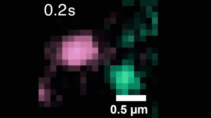 A microscopy video shows upconverting nanoparticles tagged to EGFR receptors (labeled pink and green), which track individual receptors as they dimerize. Image courtesy of the researchers.
When the EGFR molecules carried cancer-related mutations, the dimers became more stable, with the more stabilizing mutations linked to more potent cancers in people. In addition, the mutated receptors could form stable dimers even without an external stimulus prompting them to dimerize. The finding helps explain how EGFR mutations can lead to uncontrolled cell growth and cancer, and could inform efforts to target this process therapeutically.
The team discovered several other new and surprising details about how HER2 and HER3 form stable pairings with themselves, which helps illuminate the role of these molecules in related cancers.
When the research team tagged all three receptor types in one experiment, they observed a vibrant scene with receptors navigating the cell surface, finding partners, unpairing, and then finding new partners, over and over again.
Beyond shedding light on EGFR biology, the scientists hope that collaborators in other fields will apply their method to ask new scientific questions about other proteins of interest. “We think this technique could be transformative for studying molecular biology, because it enables dynamic biological processes to be observed with high spatiotemporal resolution over unprecedented timescales,” says Peng.
They are also planning to explore the method’s use in studying the mechanism of drug action, to reveal how potential therapeutics alter individual molecules over time. In addition, they will continue to improve their methods, such as making the probes smaller, brighter, and able to emit more colors.
Language development in the brainThe brain’s language network is still evolving in adolescence. But by age 4, language processing is already handled by the left side of the brain, new research finds.The brain’s capacity to use and understand language expands rapidly in the first years of life, as babies start to make sense of the words they hear and eventually begin to piece together sentences of their own. The language-processing parts of the brain that make this possible continue to evolve in older children, as they expand their vocabularies and learn to use language more flexibly.
MIT brain researchers have captured snapshots of the developing language-processing network in brain scans of hundreds of children and adolescents. Their data, reported May 16 in the journal Nature Communications, show that the network continues to mature, becoming better integrated and increasingly responsive until around age 16. But they also found that a key feature of the adult language network is established early on: its localization in the left side of the brain.
Language lateralization
It is well known that using language is mostly the job of the left hemisphere. As adults, we call on the language-processing regions there when we read, write, speak, or listen to others talk. But there was some question as to whether this left lateralization is established early in life, or might instead emerge as the language network matures, with both sides of the brain contributing to language in childhood.
To find out, researchers needed to see young brains in action — and several MIT labs had collected exactly the right kind of data. Groups led by Evelina Fedorenko, an associate professor of brain and cognitive sciences; John Gabrieli, the Grover Hermann Professor of Health Sciences and Technology; and Rebecca Saxe, the John W. Jarve (1978) Professor of Brain and Cognitive Sciences, teamed up to share brain scans from children, adolescents, and adults and compare how their brains responded to language. Fedorenko, Gabrieli, and Saxe are also investigators at the McGovern Institute for Brain Research.
In studies aimed at better understanding a variety of cognitive functions and developmental disorders, the three teams had all collected functional MRI data while subjects participated in “language localizer” tasks — an approach the Fedorenko lab developed to map the language-processing network in a person’s brain. By monitoring brain activity with functional MRI as people engage in both language tasks and non-linguistic tasks, researchers can identify parts of the brain that are exclusively dedicated to language processing, whose precise anatomic location varies across individuals.
To activate the language network, the researchers had children listen to stories inside the MRI scanner. Depending on their age, some heard excerpts of “Alice in Wonderland,” some listened to podcasts and TED talks, and others heard shorter, simpler stories. To watch their brains during a non-linguistic task, the researchers had the children listen to nonsense words.
Across the data from the three labs, which included children between the ages of 4 and 16, as well as adults for comparison, the team saw clear developmental changes in the brain’s response to language. “The integration of the system — how well different subregions of the system correlated with each other and worked together during language processing — was stronger in older children as compared to younger children,” says Ola Ozernov-Palchik, a research scientist in Gabrieli’s lab and a research assistant professor at Boston University. The system was also more strongly activated by language in older children, which may reflect their growing comprehension of what they hear.
But strikingly, almost all language processing happened on the left side of the brain, even in the youngest subjects. “From age 4 on, it seems just as lateralized as in an adult,” Gabrieli says.
Language and developmental disorders
The researchers say this finding has implications for understanding developmental conditions that impact language, including autism and dyslexia. The right side of the brain frequently gets more involved in language processing in people with these conditions than it does in typically developing children. “Almost every single developmental disorder that’s associated with language has a theory that’s related to language lateralization,” Ozernov-Palchik says.
The reason for more bilateral language processing in some disorders is debated. One idea has been that some people might use both sides of their brain for language processing because their brains are less mature. If the right side of the brain processes language early in life, scientists had reasoned, it might simply continue to do so for longer in people with autism or dyslexia than it does in neurotypical individuals. But if most people use the left side of their brains for language even when they are young, the difference can’t be attributed to a developmental delay. Other developmental differences might cause bilateral language processing instead.
The researchers don’t have the full picture yet; they still need to know what parts of the brain process language in children younger than 4. Likewise, they would like to know what the brain areas that become the language network are doing in the first months of life, when infants aren’t using language yet. They are eager to find out, both to understand fundamentals of brain development and to shed light on developmental disorders. “I think understanding that normal trajectory is really critical for interpreting what a deviation from that trajectory is,” says Amanda O’Brien, a former graduate student in Gabrieli’s lab who is now a postdoc at Harvard University.
One reason people thought lateralization might develop gradually is because damage to the left hemisphere of the brain impacts language abilities differently, depending on when it occurs. “If you have damage to the left hemisphere as an adult, you’re very likely to end up with some form of aphasia, at least temporarily,” Fedorenko explains. “But a lot of the time, with early damage to the left hemisphere, you grow up and you’re totally fine. The language can just develop in the right hemisphere.”
Some scientists suspected that the right side of the brain was able to take over language processing in children who suffered early-life brain damage because it was already participating in this function at the time. But the team’s findings suggest the developing brain may be nimbler than that. “Our data tell you that this early plasticity apparently happens in spite of the fact that by age 4, we see these very strongly lateralized responses already,” Fedorenko says.
Two from MIT named 2026 Knight-Hennessy ScholarsThe prestigious fellowship funds graduate studies at Stanford University.MIT master’s student Sunshine Jiang ’25 and Rupert Li ’24 are recipients of this year’s Knight-Hennessy Scholarship. Now in its ninth year, the highly competitive scholarship provides up to three years of financial support for graduate studies at Stanford University.
Sunshine Jiang ’25
Sunshine Jiang, from Hangzhou, China, graduated from MIT in 2025 with a bachelor’s degree as a double major in physics and electrical engineering and computer science, along with minors in mathematics and economics. She will receive her master of engineering degree this month and will start her PhD in computer science at Stanford School of Engineering this fall.
Jiang researches embodied artificial intelligence and robotics, developing data-efficient, adaptive systems for general-purpose robots that broaden accessibility. She has presented her research at major conferences, including the Conference on Robot Learning, the International Conference on Robotics and Automation, and the International Conference on Learning Representations.
Jiang led the development of AI-powered systems that provide access to traditional Chinese art in rural classrooms, founded cross-country programs that expand girls’ access to STEM education, and created a Covid-19 documentary amplifying community voices, which was featured on China Daily.
Rupert Li ’24
Rupert Li, from Portland, Oregon, is currently pursuing a PhD in mathematics at Stanford School of Humanities and Sciences. He graduated from MIT in 2024 with a bachelor’s degree, double majoring in mathematics and computer science, economics, and data science. Along with his bachelor’s degree, he also received a master’s degree in data science. Li then traveled to the United Kingdom as a Marshall Scholar, where he earned a master’s degree in mathematics from the University of Cambridge.
Li’s research interests lie in probability, discrete geometry, and combinatorics. He enjoys serving as a mentor for MIT PRIMES-USA, a high school math research program, and previously served as an advisor for the Duluth REU, an undergraduate math research program. In addition to the Knight-Hennessy Scholarship and the Marshall Scholarship, he has been awarded the Hertz Fellowship, P.D. Soros Fellowship, and the Goldwater Scholarship, and he received honorable mention for the Frank and Brennie Morgan Prize.
A new way to spot signs of dark matterGravitational waves emitted by colliding black holes may bear imprints of dark matter, which physicists could detect with a new model.Dark matter is thought to make up most of the matter in the universe, but the only way it interacts with its surroundings is through gravity. If two colliding black holes spiral through a dense region of dark matter and merge, gravitational waves rippling across space and time could carry an imprint of that dark matter.
Now, physicists may be able to spot such imprints of dark matter in gravitational waves that are detected on Earth.
Researchers at MIT and in Europe have developed a method that makes predictions for what a gravitational wave should look like if it were produced by black holes that moved through dark matter, rather than empty space. They applied the technique to publicly available gravitational-wave data previously recorded by LIGO-Virgo-KAGRA (LVK), the global network of observatories that detect gravitational waves from black hole mergers and other far-off astrophysical sources.
The researchers looked through the gravitational-wave signals recorded over the LVK’s first three observing runs. From 28 of the clearest signals, the team found that 27 originated from black holes that merged in a vacuum, as physicists expected. But the pattern of one signal, GW190728, showed possible signs of a dark matter imprint.
The scientists emphasize that they have not detected dark matter. Rather, the new method offers a new way to screen gravitational-wave data for hints of dark matter, which physicists can then follow up and confirm with other techniques.
“We know that dark matter is around us. It just has to be dense enough for us to see its effects,” says Josu Aurrekoetxea, a postdoc in the MIT Department of Physics. “Black holes provide a mechanism to enhance this density, which we can now search for by analyzing the gravitational waves emitted when they merge.”
Aurrekoetxea and his colleagues report their results in a study appearing today in Physical Review Letters. The study’s co-authors are LVK member Soumen Roy of Université Catholique de Louvain (UCLouvain) in Belgium, Rodrigo Vicente of the University of Amsterdam, Katy Clough of Queen Mary University of London, and Pedro Ferreira of Oxford University.
A dark pull
Dark matter is an invisible, hypothetical form of matter that, unlike normal everyday matter, has no interactions with the electromagnetic force. Dark matter can pass through light, magnetic fields, and any other form of energy along the electromagnetic spectrum without leaving a trace. The only evidence that dark matter exists is through its apparent interaction with one other force: gravity.
By observing how gravity bends around distant galaxies, astronomers have surmised that there must be an extra force, outside of the galaxies’ own gravitational pull, to explain the bending fields, or “lensing.” This extra force, physicists suspect, is dark matter, which could account for over 85 percent of the matter in the universe. But exactly what dark matter is is a matter of huge debate, with theories for dark matter particles that range widely in particle size and properties.
One class of proposed dark matter consists of “light scalar” particles, whose masses are many orders of magnitude lighter than an electron. Theorists predict that such dark matter should behave not just as particles, but also as coordinated waves when moving near black holes.
When waves of dark matter come in contact with a rapidly spinning black hole, physicists predict that the black hole's rotational energy can be transferred to the dark matter, amplifying it. This phenomenon, known as superradiance, would whip up the waves to extremely high densities of dark matter, akin to churning cream into butter.
At high enough densities, light scalar dark matter, which is invisible by all other accounts, should leave an imprint on the gravitational waves that reverberate from the colliding black holes.
But exactly what would that imprint look like? And could such an imprint be detectable in gravitational waves that arrive on Earth, from black holes that merged many millions of light years away?
For answers to those questions, Aurrekoetxea and his colleagues developed a model to predict the gravitational waveform, or the pattern of gravitational waves that two black holes would produce, if they collided in an environment of dark matter, versus in a vacuum (empty space, with no dark matter).
An imprint’s prediction
For their new study, the team performed detailed numerical simulations to predict the gravitational wave that would be produced given various properties of two colliding black holes — a system known as a “black hole binary.” They considered black hole binaries across a range of scenarios and properties, for example, varying the size and mass of each black hole, the environment of dark matter that the black holes might pass through, and the density of the dark matter that the black holes would spin up.
They designed the model to predict what a gravitational wave from a black hole binary would look like if it carried an imprint of dark matter, and furthermore, what that wave would look like if it traveled a given distance across space and time, to eventually arrive at a detector on Earth.
With their model, they looked to see whether any gravitational-wave signals that have been detected on Earth match their predicted patterns of dark matter imprints. To do so, they applied the model to publicly-available data recorded by LVK over the observatories’ first three observing runs. The observatories have picked up hundreds of gravitational-wave signals during this period. For their purposes, the researchers focused on the clearest signals, comprising gravitational waves from 28 separate events.
For each event, the team compared the pattern of the actual gravitational wave against their model of what the signal would look like if it were generated by the same event in an environment of dark matter. They also compared the gravitational wave to the more expected scenario in which the signal was produced in a vacuum.
Of the 28 clearest signals that they analyzed, 27 were solidly within the predictions for having been produced in a vacuum. However, the pattern of one event, GW190728, showed a “preference,” or an agreement with the team’s dark matter model. In other words, the signal may carry an imprint of dark matter.
GW190728 is a gravitational wave that is named after the date that it was detected — on July 28, 2019. Scientists previously determined that the gravitational wave originated from a black hole binary with a total mass of about 20 times the mass of the sun. With their model, the team showed that such a system could have merged through a dense cloud of dark matter and produced a similar gravitational wave to GW190728.
“The statistical significance of this is not high enough to claim a detection of dark matter, and further checks should be performed by independent groups,” Aurrekoetxea says. “What we think is important to highlight is that without waveform models like ours, we could be detecting black hole mergers in dark matter environments, but systematically classifying them as having occurred in vacuum.”
“We now have the potential to discover dark matter around black holes as the LVK detectors keep collecting data in the coming years,” says co-author Soumen Roy, who led the data analysis part of the work. “It is an exciting time to search for new physics using gravitational waves.”
“Using black holes to look for dark matter would be fantastic,” adds co-author Rodrigo Vicente, who developed the analytical model of the signal. “We would be able to probe dark matter at scales much smaller than ever before.”
This work was supported, in part, by the U.S. National Science Foundation and MIT’s Center for Theoretical Physics — a Leinweber Institute.
Powerful shrinking technique could enable devices that compute with lightMIT researchers created tiny 3D photonic devices with features small enough to channel visible light.Using a new technique that can create vacancies at any site across a material and then shrink it to about 1/2,000 of its original volume, MIT researchers have designed nanotechnology devices that could be used for optical computing and other applications involving the manipulation of visible light.
The new fabrication technique, known as “implosion carving,” allows researchers to imprint features throughout a hydrogel using photopatterning. If patterned with a resolution of about 800 nanometers, these features can then be shrunk to less than 100 nanometers.
Because that resolution is smaller than the wavelength of light, the devices can bend light in specific ways that allow them to perform optical computations.

“In order to enable nanophotonic applications in visible light, we need to make nanostructures with feature sizes with a resolution less than 100 nanometers. Only in that way can we precisely create the structure that can manipulate visible light,” says Quansan Yang, a former MIT postdoc, now an assistant professor at the University of Washington, and one of the lead authors of the new study.
In their paper, the researchers demonstrated a photonic device that can perform a simple digit-classification task, but future versions could be used for high-speed imaging and information processing, they say.
Gaojie Yang, a former MIT postdoc, is the co-lead author of the paper, which appears today in Nature Photonics. The paper’s senior authors are Peter So, director of the MIT Laser Biomedical Research Center (LBCR) and an MIT professor of biological engineering and mechanical engineering, and Edward Boyden, the Y. Eva Tan Professor in Neurotechnology at MIT and a professor of biological engineering, media arts and sciences, and brain and cognitive sciences. Boyden is also a Howard Hughes Medical Institute investigator and a member of MIT’s McGovern Institute for Brain Research, the Yang Tan Collective, and Koch Institute for Integrative Cancer Research.
Nanoscale feature sizes
Photonic devices, which transmit and manipulate light, hold potential for use as optical computer chips that could offer an energy-efficient alternative to semiconductor chips. However, existing techniques for creating 3D photonic devices haven’t yet achieved the 100-nanometer resolution that is needed to channel visible light, which has wavelengths between 380 and 750 nanometers.
Using an additive manufacturing technique called two-photon lithography, researchers can use light to create 3D nanoscale features, but with a resolution larger than 100 nanometers. Another technique, known as electron-beam lithography, can be used to etch smaller-resolution features onto a silicon chip, but it doesn’t generate 3D structures.
To make 3D devices with the necessary feature size, the researchers extended the concept of “implosion fabrication,” which Boyden’s lab developed in 2018, to create a new variant called “implosion carving.” In implosion carving, a laser creates vacancies — tiny voids where the hydrogel material has been removed — at precisely targeted locations. These vacancies exhibit different optical properties than the surrounding hydrogel. The hydrogel is then shrunk to bring the patterned features down to the nanoscale.
The carving process begins with immersing the hydrogel in a photosensitizing dye. Then, the researchers use a laser to excite the photosensitizer at specific places in the gel, which in turn generates reactive oxygen species that cut the bonds holding the hydrogel together. This creates a vacancy in that spot.
Once the desired vacancy pattern has been carved into the hydrogel, the researchers shrink it using a two-step process. First, they soak it in a solution containing ions, which causes it to shrink about tenfold in each dimension. To shrink it a little more, and to remove the watery solution, the hydrogel then undergoes a process called supercritical drying, which can remove liquid from a gel without damaging it.
At the end of the process, the hydrogel has been shrunk more than tenfold in each dimension, leading to a 2,000-fold reduction in volume.
Computing with light
To demonstrate the versatility of this technique, the researchers used it to create several 3D shapes, including a helix and a structure inspired by a butterfly wing. Some of these structures are too thin, and have too high an aspect ratio, to be stably created using conventional two-photon lithography.
The researchers also created a device that could perform a simple calculation known as digit classification, a task that is traditionally used to test the performance of neural networks. During this task, the device was presented with a digit, such as 1 or 5, and had to light up a specific location to indicate which number was detected.
To achieve this, the researchers patterned vacancies throughout the device so that it would act like a neural network. The pattern of vacancies would diffract input light as it passed through many layers of patterned hydrogel, so that the output light was determined by the shape of the digit that was entered into the system.
“This is a purely optical system that effectively performs optical computing,” So says.
“One of the very attractive features of this technology is that you can manipulate the property of the material at every tiny location,” says Dushan Wadduwage, an assistant professor at Old Dominion University and former MIT postdoc, who is also an author of the paper. “You have millions of different locations that you need to decide the property of, and that turns into a really interesting design problem where we can use deep-learning algorithms to find designs over these millions of parameters and come up with parts that go into optical systems in new ways.”
The researchers now plan to use the same principles to build optical devices that could classify cells based on their state as they flow through a microfluidic device. This could help identify rare cells such as circulating tumor cells in a blood sample, they say.
This approach could also enable the creation of high-throughput imaging techniques for applications such as analyzing tissue samples from biopsies or surgical specimens. And, if adapted to work with other materials such as hydrophobic polymers, it could also be used to create channels within 3D nanofluidic devices.
Other authors of the paper include Gaojie Yang, Takahiro Nambara, Hiroyuki Kusaka, Yuichiro Kunai, Alex Matlock, Corban Swain, Brett Pryor, Yannick Salamin, Daniel Oran, Hasindu Kariyawasam, Ramith Hettiarachchi, and Marin Soljacic.
The research was funded, in part, by the MIT-Fujikura Partnership Fund, the U.S. Army Research Office through the Institute for Soldier Nanotechnologies at MIT, Lisa Yang and Y. Eva Tan, John Doerr, the Open Philanthropy Project, the Howard Hughes Medical Institute, and the U.S. National Institutes of Health.
Improving the reliability of circuits for quantum computersA new technique helps scientists measure a phenomenon that can cause quantum circuits to perform differently than expected, increasing the error in computations.Quantum computers could someday solve pressing problems that are too convoluted for classical computers, such as modeling complex molecular interactions to streamline drug discovery and materials development.
But to build a superconducting quantum computer that is large and resilient enough for real-world applications, scientists must precisely engineer thousands of quantum circuits so they perform operations with the lowest possible error rate.
To help scientists design more predictable circuits, researchers from MIT and Lincoln Laboratory developed a technique to measure a property that can unexpectedly cause a superconducting quantum circuit to deviate from its expected behavior. Their analysis revealed the source of these distortions, known as second-order harmonic corrections, leading to underperforming circuit architectures.
The MIT researchers fabricated a device to detect second-order harmonic corrections, identify their origin, and precisely measure their strength. This technique could help scientists deliberately design quantum circuits that can counteract the effects of these deviations.
This is especially important in larger and more complicated quantum circuits, where the negative impact of second-order harmonic corrections can be amplified.
“As we make our quantum computers bigger and we want to have more precise control over the parameters of these devices, identifying and measuring these effects is going to be important for us to have a precise understanding of how these systems are constructed. It is always important to keep diving down into the circuit to see if there is an effect you didn’t expect, which impacts how your device is performing,” says Max Hays, a research scientist in the Engineering Quantum Systems (EQuS) group of the Research Laboratory of Electronics (RLE) and co-lead author of a paper on this research.
Hays is joined on the paper by co-lead author Junghyun Kim, an electrical engineering and computer science (EECS) graduate student in the EQuS group; senior author William D. Oliver, the Henry Ellis Warren (1894) Professor of EECS and professor of physics, leader of the EQuS group, director of the Center for Quantum Engineering, and associate director of RLE; as well as others at MIT and Lincoln Laboratory. The research appears today in Nature Physics.
A pair-wise problem
In a quantum computer that utilizes superconducting circuits, which is one of many potential computing platforms, Josephson junctions are critical elements that enable the transfer and manipulation of information. These devices utilize two superconducting wires that are brought very close together, with a nanometer-scale barrier between them. Like a traditional circuit, the electric charge in Josephson junctions is carried by electrons.
But in a superconducting circuit, charge-carrying electrons pair up, forming what are called Cooper pairs. These Cooper pairs can “quantum tunnel” through the barrier between the two wires, transporting current from one wire to the other.
Cooper pairs can usually only tunnel one pair at a time, which is a key property that makes quantum computation possible.
“If you try to force more Cooper pairs through, it just doesn’t work. This non-linear effect is extremely important for all our circuits. If we didn’t have that effect, then we wouldn’t be able to control or manipulate any quantum information that we store in these circuits,” Hays explains.
But sometimes, Cooper pairs can unexpectedly squeeze through the barrier two at a time, an effect that is known as a second-order harmonic correction. This effect limits the performance of a quantum circuit that has been configured to only allow single-pair tunneling.
“If two Cooper pairs tunnel at the same time, then the assumption we used to build our circuit doesn’t apply anymore. We need to fix the circuit so it can handle that,” Kim says.
But before they can fix the circuit, scientists need to know the source and strength of these distortions.
To obtain this information, the MIT researchers fabricated a quantum circuit so it would be very sensitive to these effects. Essentially, the device is designed to suppress the quantum tunneling process of single Cooper pairs, while allowing the two-pair tunneling process to continue.
In this way, they can detect the presence of second-order harmonic corrections and precisely measure their strength.
Straight to the source
They can also use this circuit to pinpoint the source of these harmonics, which helps researchers identify the best way to correct for them.
There are two potential sources of second-order harmonics — one source is intrinsic to the dynamics of the Josephson junction and the other is caused by the wires connecting the junction to other circuit elements.
While prior research had indicated the second-order harmonics could be due to the dynamics of the junction, the MIT researchers found that additional inductance — the tendency to oppose changes in the flow of electric current —from wires in the circuit was the actual source in their devices.
“This is important because, if we know where the second-order harmonic correction is coming from, we can predict how strong it is likely to be, and use that information to engineer more predictable circuits that will hopefully perform better,” Hays says.
In the future, the researchers want to design experiments that more accurately predict how a device will perform when second-order harmonic corrections occur. They also want to study other sources of second-order harmonic corrections and whether those sources could have negative impacts on a circuit under different fabrication conditions.
This work is funded, in part, by the U.S. Department of Energy, the U.S. Co-design Center for Quantum Advantage, the U.S. Air Force, the Korea Foundation for Advanced Studies, and the Intelligence Community Postdoctoral Research Fellowship Program at MIT.
Astronomers pin down the origins of a planetary odd coupleNew measurements of a hot Jupiter and its mini-Neptune companion suggest both planets formed surprisingly far away from their host star.Across the Milky Way galaxy, a planetary odd couple is circling a star some 190 light years from Earth. A normally “lonely” hot Jupiter is sharing space with a mini-Neptune, in a rare and unlikely pairing that’s had astronomers puzzled since the system’s discovery in 2020.
Now MIT scientists have caught a glimpse into the atmosphere of the mini-Neptune, which is circling inside the orbit of its Jupiter-sized companion, and discovered clues to explain the origins of this unusual planetary system.
In a study appearing today in Astrophysical Journal Letters, the scientists report on new measurements of the mini-Neptune’s atmosphere, made using NASA’s James Webb Space Telescope (JWST). It is the first time astronomers have measured the composition of a mini-Neptune that resides inside the orbit of a hot Jupiter.
Their measurements reveal that the smaller planet has a “heavy” atmosphere that is rich with water vapor, carbon dioxide, sulfur dioxide, and hints of methane. Such a heavy atmosphere would not have been acquired by the planet if it had formed in its current location, very close to its star.
Instead, the scientists say their findings point to an alternate origin story: Both the mini-Neptune and the hot Jupiter may have formed much farther away, in the colder region of the protoplanetary disk. There, the planets could slowly build up atmospheres of ice and other volatiles. Over time, the planets were likely drawn in toward the star in a gradual process that kept them close, with their atmospheres intact.
The team’s results are the first to show that mini-Neptunes can form beyond a star’s “frost line.” This boundary refers to the minimum distance from a star where the temperature is low enough that water instantly condenses into ice.
“This is the first time we’ve observed the atmosphere of a planet that is inside the orbit of a hot Jupiter,” says Saugata Barat, a postdoc in MIT’s Kavli Institute for Astrophysics and Space Research and the lead author of the study. “This measurement tells us this mini-Neptune indeed formed beyond the frost line, giving confirmation that this formation channel does exist.”
The team consists of astronomers around the world, including Andrew Vanderburg, a visiting assistant professor at MIT, and co-authors from multiple other institutions including the Harvard and Smithsonian Center for Astrophysics, the University of South Queensland, the University of Texas at Austin, and Lund University.
A “one-of-a-kind” system
As their name implies, mini-Neptunes are planets that are less massive than Neptune. They are considered to be gas dwarfs, which are made mostly of gas, with an inner, rocky core. Mini-Neptunes are the most commonly found planet in the Milky Way, though, interestingly, no such world exists in our own solar system. Astronomers have observed many planets circling a wide variety of stars in a range of planetary systems. Mini-Neptunes, then, are generally considered to be garden-variety planets.
But in 2020, Chelsea X. Huang, then a Torres Postdoctoral fellow at MIT (now on the faculty at University of South Queensland), discovered a mini-Neptune in a rare and puzzling circumstance: The planet appeared to be circling its star with an unlikely companion — a hot Jupiter.
The astronomers made their discovery using NASA’s Transiting Exoplanet Survey Satellite (TESS). They analyzed TESS’ measurements of TOI-1130, a star located 190 light years from Earth, and detected signs of a mini-Neptune and a hot Jupiter, orbiting the star every four and eight days respectively.
“This was a one-of-a-kind system,” says Huang. “Hot Jupiters are ‘lonely,’ meaning they don’t have companion planets inside their orbits. They are so massive, and their gravity is so strong, that whatever is inside their orbit just gets scattered away. But somehow, with this hot Jupiter, an inner companion has survived. And that raises questions about how such a system could form.”
A spot-on snapshot
The 2020 discovery of TOI-1130 and its odd planetary pair inspired Huang, Vanderburg, and their colleagues to take a closer look at the planets, and specifically, their atmospheres, with JWST. In its new study, the team reports its analysis of TOI-1130b — the inner-orbiting mini-Neptune.
Catching the planet at just the right time was their first challenge. Most planets circle their star with a regular, predictable period, like the tick of a clock. But the mini-Neptune and the hot Jupiter were found to be in “mean motion resonance,” meaning that each can affect the other’s motion, pulling and tugging, and slightly varying the time each takes to orbit their star. This made it tricky to predict when JWST could get a clear view.
The team, led by Judith Korth of Lund University, assembled as many past observations of the system as they could, and developed a model to predict when each planet would pass by the star at an angle that JWST could observe.
“It was a challenging prediction, and we had to be spot-on,” Barat says.
In the end, the team was able to catch a direct and detailed snapshot of both planets.
“The beauty of JWST is that it does not observe just in one color, but at different colors, or wavelengths,” Barat explains. “And the specific wavelengths that a planet absorbs can tell you a lot about the composition of its atmosphere.”
From JWST’s measurements, the team found that the planet absorbed wavelengths specifically for water, carbon dioxide, sulfur dioxide, and to a lesser degree, methane. These molecules are heavier than hydrogen and helium, which constitute lighter atmospheres. Astronomers had assumed that, if mini-Neptunes formed very close to their star, they should have light atmospheres.
But the team’s new results counter that assumption and offer a new way that mini-Neptunes could form. Since heavier molecules were found in the atmosphere of TOI-1130b, which resides very close to its star, the scientists say the only possible explanation for its composition is that the planet formed much farther out than its current location.
The planet likely accumulated its heavy atmosphere of water and other volatiles such as carbon dioxide and sulfur dioxide in the icy region beyond the star’s frost line. In this much colder environment, water condenses onto bits of dust to form icy pebbles, which an infant planet can draw into its atmosphere. The water evaporates as it slowly migrates in closer to its star.
Barat says the team’s detection of heavy molecules in the atmosphere of TOI-1130b confirms that the planet — and likely its hot Jupiter companion — formed in the outskirts of the system. Through gradual migration, the two planets would be able to stay close together and keep their atmospheres intact.
“This system represents one of the rarest architectures that astronomers have ever found,” Barat says. “The observations of TOI-1130b provide the first hint that such mini-Neptunes that form beyond the water/ice line are indeed present in nature.”
This work was supported, in part, by NASA.
Biologist Joey Davis explores how cells build complex structures His studies have shed light on the assembly instructions that govern ribosomes, the critical protein-building machines of the cell.Ribosomes, the cellular machines that assemble proteins, are made from dozens of proteins and RNA molecules. Putting all of those pieces together is a complex puzzle — one that MIT Associate Professor Joey Davis PhD ’10 revels in trying to solve.
Understanding how these structures form and later break down could help researchers learn more about how disruptions of these fundamental processes can lead to disease. But, as Davis points out, it’s also an interesting biological question.
“Our long-term goal is to really understand how the natural world assembles these huge complexes rapidly and efficiently. It’s a fundamentally interesting question to think about how these things get put together,” he says.
His work has helped reveal that unlike building a house, which happens in a prescribed sequence of steps — pouring the foundation, building the frame, putting on the roof, then doing electrical and plumbing work — ribosomes can be assembled in a more flexible way. Cells can even skip an assembly step and then come back to it later.
“In these natural systems, it seems like the assembly pathways are much more dynamic and flexible,” he says. “It appears that evolution has selected pathways that aren’t strictly ordered in the way we would think about an assembly line, where you always put in one component, then the next, and then the next. We’re excited to understand the selective advantages of such approaches.”
A love of discovery
Davis’ interest in how things are put together developed early in life, inspired by his father, a carpenter who framed houses. During the mid-1980s, the family moved from Colorado to Southern California, where his father worked in construction during a housing boom there.
“I was always interested in building things, which I think probably came from being around my dad and other builders,” Davis says.
As an undergraduate at the University of California at Berkeley, where he majored in computer science and biological engineering, Davis’ interests turned toward smaller scales, in the realm of cells and molecules. During his junior year, he started working in the lab of chemistry professor Michael Marletta, who studies molecular-level biological interactions.
In the lab, Davis investigated how enzymes that contain heme are able to preferentially bind to either oxygen or nitric oxide, two gases that are very similar in structure. That work kindled a love of studying the natural world and pursuing discoveries in fundamental science.
“Being in the Marletta lab and seeing students and postdocs that were really passionate about these problems had a big impact on me,” Davis says. “The goal was to understand the fundamentals of how molecular discrimination works, and the idea of discovery for the sake of discovery was thrilling.”
After graduating from Berkeley, Davis spent another year working in Marletta’s lab, and then a year working odd jobs, before heading to MIT to pursue a PhD in biology. There, he worked with Professor Bob Sauer, now emeritus, who studied the relationship between protein structure and function, with a particular focus on the molecular machines that degrade or remodel proteins.
Davis’ thesis research centered on enzymes called AAA proteases, which remove damaged proteins from cellular membranes and send them to cell organelles that break them down. In addition to studying the structure and function of the proteases, Davis worked on ways to engineer them to tag specific proteins for destruction.
That work led him into synthetic biology, which he used to develop genetic parts that drive production of proteins of interest. Some of those parts ended up being used by the biotech startup Ginkgo Bioworks, where Davis took a job as a senior scientist after graduating.
Working at Ginkgo Bioworks allowed Davis to stay in Boston while his partner finished her PhD. The couple then moved back to California, where Davis worked as a postdoc at Scripps Research, which was home to one of the first direct electron detection cameras for cryo-electron microscopy (cryo-EM). These detectors allow researchers to generate structures with near atomic resolution. At Scripps, Davis began using them to study ribosomes as they were being assembled.
Peering into the ribosome
After joining the MIT faculty in 2017, Davis continued his work on ribosomes and assembled a lab group that includes students from a variety of backgrounds who work together to develop new ways to explore biological phenomena.
“I have a mix of method developers and biologists in the group, and the work from each of them informs each other,” Davis says. “My lab goes back and forth between building sets of tools to answer biological questions, and then as we’re answering those questions, it motivates the next generation of tool development.”
During ribosome assembly, RNA molecules fold themselves into the correct shapes, creating docking sites for proteins to attach. Then, more RNA molecules come in and fold themselves into the structure.
“It’s a beautifully coupled process by which the cell folds hundreds of RNA helices and binds on the order of 50 proteins, and it does it in two minutes from start to finish. E. coli does this 100,000 times per hour, and it’s amazing how rapid and efficient the process is,” Davis says.
Cryo-EM allows scientists to capture this process in minute detail. It can be used to take hundreds of thousands of two-dimensional images of ribosome samples frozen in a thin layer of ice, from different angles. Computer algorithms then piece together these images into a three-dimensional representation of the ribosome.
To gain insight into how ribosomes are assembled, researchers can stall the process at different points and then analyze the resulting structures. In 2021, Davis’s lab developed a new method called CryoDRGN, which uses neural networks to analyze cryo-EM data and generate the full ensemble of structures that were present in the sample.
This work has shown that when certain steps of ribosome assembly are blocked, many different structures result, suggesting that the assembly can occur in a variety of ways.
In future work, Davis aims to dramatically increase the throughput of cryo-EM to generate datasets of protein structures that could help improve the AI-based models that are now used to predict protein structures.
“There are still huge swaths of sequence space that these models are very poor at predicting, but if we could collect data on those sequences en masse, that could potentially serve as key training data for a next-generation protein structure prediction method that could fill out that space,” he says.
Beacon Biosignals is mapping the brain during sleepFounded by Jake Donoghue PhD ’19 and former MIT researcher Jarrett Revels, the company is creating an AI-driven platform to help diagnose and treat disease.The human brain remains one of the most fascinating and perplexing mysteries in medicine. Scientists still struggle to match neurological activity with brain function and detect problems early, slowing efforts to treat neurological disorders and other diseases.
Beacon Biosignals is working to make sense of the brain by monitoring its activity while people sleep. The company, which was founded by Jake Donoghue PhD ’19 and former MIT researcher Jarrett Revels, developed a lightweight headband that uses electroencephalogram (EEG) technology to measure brain activity while people enjoy their normal sleep routines at home. Those data are processed by machine-learning algorithms to monitor the effects of novel treatments, find new signs of disease progression, and create patient cohorts for clinical trials.
“There’s a step-change in what becomes possible when you remove the sleep lab and bring clinical-grade EEG into the home,” says Donoghue, who serves as Beacon’s CEO. “It turns sleep from a constrained, facility-based test into a scalable source of high-quality data for diagnostics, drug development, and longitudinal brain health.”
Beacon partners with pharmaceutical companies to accelerate its path to patients. The company’s FDA 510(k)-cleared medical device has already been used in over 40 clinical trials across the globe as part of studies aimed at treating conditions including major depressive disorder, schizophrenia, narcolepsy, idiopathic hypersomnia, Alzheimer’s disease, and Parkinson’s disease.
With each deployment, Beacon learns more about how the brain works — insights it is using to create a “foundation model” of the brain.
“It’s our belief that the dataset that’s going to transform brain health doesn’t exist yet — but we are rapidly creating it,” Donoghue says. “Our platform can characterize the heterogeneity of disease progression, generating dynamic insights that are impossible to fully capture through static modalities like sequencing or imaging. The brain is an electric organ and changes through synaptic plasticity, so tracking brain function across many diseases at scale will allow us to discover novel subgroups of diseases and map them over time.”
Illuminating the brain
Donoghue trained in the Harvard-MIT Program in Health Sciences and Technology, conducting clinical training for an MD while completing his PhD in neuroscience at MIT under the guidance of Earl Miller, MIT's Picower Professor in Brain and Cognitive Sciences and The Picower Institute for Learning and Memory. While in the program, Donoghue trained at Massachusetts General Hospital and Boston Children’s Hospital, where he helped care for patients, including in oncology, during the rise of genomic sequencing to guide precision cancer therapies. He later worked in neurology and psychiatry, where care often relied on more iterative approaches — highlighting an opportunity to bring similarly data-driven precision to brain health.
“What struck me most was the inability to measure brain function in the ways that cardiologists can longitudinally monitor cardiac function in patients from home,” Donoghue says. “At MIT, I built this conviction that processing a lot of brain data and working to correlate that with brain function would be transformative to how these neurological diseases are identified and treated.”
Toward the end of his training, Donoghue began developing his ideas further, engaging with mentors including HST and Harvard Medical School professors Sydney Cash and Brandon Westover. He had met Revels, who was working as a research software engineer in MIT’s Julia Lab, during his PhD, and convinced him to co-found Beacon with him in 2019.
“We decided building a business to understand the organ of interest — the brain — would be a great start to understanding heterogeneous neuropsychiatric diseases and building better treatments,” Donoghue recalls.
Beacon began as a computation and analytics company building wearable devices to expand clinical impact and reach. From its early days, Beacon has been partnering with large pharmaceutical companies running clinical trials, offering a less invasive way to watch brain activity and learn how their drugs are impacting the brain as well as how patients sleep.
“It was clear sleep was the right window to understand the brain,” Donoghue says. “Neural activity during sleep can be an order of magnitude higher and more structured, almost like a language. It’s a great surface area for understanding brain function and how different drugs affect the brain.”
Donoghue says Beacon’s devices can collect lab-grade data on each patient for multiple sequential nights, resulting in higher quality assessment. The company uses machine learning to extract insights, such as the time patients spend in different sleep stages and the number of small awakenings that occur throughout the night. It can also detect subtle sleep architecture changes that might lead to cognitive decline.
“We’re starting to take features of sleep activity and link them to outcomes in a way that’s never been done with this level of precision,” Donoghue says.
To date, Beacon has taken part in clinical trials for sleep and psychiatric disorders as well as neurodegenerative diseases, where sleep changes can emerge years before the presentation of symptoms.
“We do a lot of work in areas like Alzheimer’s disease and Parkinson’s, which affected my grandfather,” Donoghue says. “We’re analyzing features of rapid-eye-movement and slow-wave sleep to detect early changes that precede clinical symptoms. It’s an opportunity to move these diseases from late recognition to much earlier, data-driven detection.”
Improving brain treatments for millions
Last year, Beacon acquired an at-home sleep apnea testing company that serves more than 100,000 patients each year across the U.S., accelerating access to high-quality, comprehensive testing in the home and expanding the reach of its platform. Then in November, the company raised $97 million to accelerate that expansion.
“The vision has always been to reach patients and help people at scale,” Donoghue says. “What’s powerful is that we’re building a longitudinal record of brain function over time,” Donoghue says. “A patient might come in for sleep apnea screening, but if they develop Parkinson’s years later, that earlier data becomes a window into the disease before symptoms emerged. That turns routine testing into a foundation for entirely new prognostic biomarkers — and a path to detecting and intervening in brain disease earlier, potentially before symptoms ever begin.”